
|
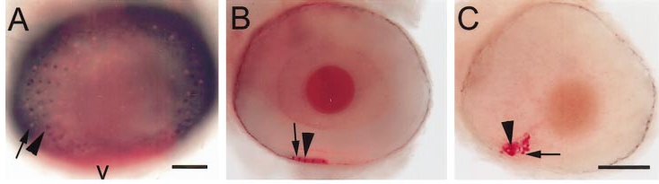
Fig. 5 Recruitment of rods and red cones in wild-type and syut4 embryos and in shh/twhh antisense-injected wild-type embryos. Eyes were from 75-hpf embryos, hybridized with GFrod (red color; arrows) and GFred (dark color; arrowheads). (A) Wild-type embryo. Image is a superimposed projection of several focal planes (using the ?apply image? function in Adobe PhotoShop), in bright-field and epifluorescence optics. Scale bar (applies to A and B), 50 μm; v, ventral. (B) Eye of embryo injected with shh/twhh antisense oligos. (C) syut4 embryo. B and C are superimposed epifluorescent and bright-field images, but from a single focal plane that contained all labeled photoreceptors. Scale bar in C, 50 μm.
Reprinted from Developmental Biology, 220(2), Stenkamp, D.L., Frey, R.A., Prabhudesai, S.N., and Raymond, P.A., Function for hedgehog genes in zebrafish retinal development, 238-252, Copyright (2000) with permission from Elsevier. Full text @ Dev. Biol.