Fig. S12
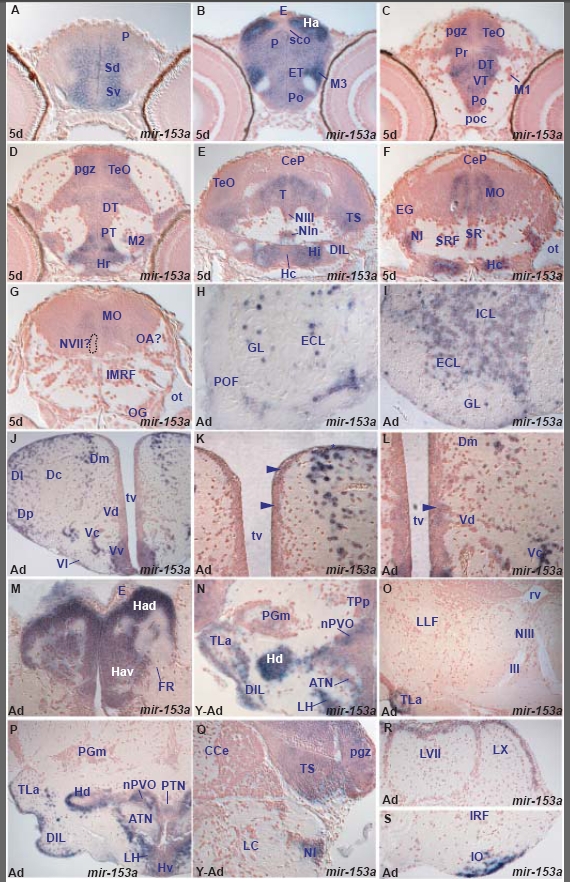
miR-153a expression in the zebrafish brain.
miR-153a shows expression in both periventricular and differentiating cells in the larval brain and has more restricted expression compared to miR-124. It is expressed in periventricular cells only in a few cases. In addition, miR-153a expression shows quantitative differences from one area to another and is conserved in many areas between larval and adult rostral brain but also presents some regional differences (Tables C,H).
Rostrally, miR-153a is expressed in both larval and adult telencephalic pallial and subpallial areas, preoptic area, habenula, dorsal and ventral thalamus, hypothalamus, posterior tuberculum, mesencephalic tectal periventricular gray zone and interpeduncular nucleus. In addition to these similarities between rostral larval and adult brain, we observe some differences: miR-153a is expressed only in the adult olfactory bulb whereas it is expressed only in the larval periventricular pretectum. Furthermore, it is expressed in the entire larval tegmentum but in the adult it is restricted in the semicircular torus. Caudally, miR-153a is expressed in the young adult and adult hindbrain, in the isthmic nucleus, nucleus of lateral valvula, perilemniscal nucleus, facial and vagal lobes and area postrema. Within the larval hindbrain we observe columns of miR-153a expression but it is difficult to establish a precise correlation with the adult nuclei. Finally, we observe quantitative differences in miR-153a expression between regions. For example miR-153a expression is particularly highly expressed in the larval habenula and migrated eminentia thalami.
A. transverse section through the larval telencephalon showing miR-153a in the ventral (Sv) and dorsal (Sd) subpallium and pallium (P).
B. transverse section through the larval caudal telencephalon and diencephalon showing miR-153a expressing cells in the preoptic area (Po), eminentia thalami (ET), migrated eminentia thalami (M3), pallium (P) and habenula (Ha).
C. transverse section through the larval diencephalon and rostral optic tectum showing miR-153a expressing cells in the preoptic area (Po), ventral thalamus (VT), dorsal thalamus (DT), periventricular pretectum (Pr) and periventricular gray zone (pgz) of the optic tectum (TeO).
D. transverse section through the larval diencephalon and midbrain showing miR-153a expressing cells in the rostral hypothalamus (Hr), periventricular (PT) and migrated (M2) posterior tubercular area, dorsal thalamus (DT) and periventricular gray zone (pgz) of the optic tectum (TeO).
E. transverse section through the larval caudal hypothalamus, midbrain and rostal hindbrain showing miR-153a expressing cells in the intermediate (Hi) and caudal (Hc) hypothalamus, diffuse nucleus of inferior lobe (DIL), interpeduncular nucleus (NIn), tegmentum (T), oculomotor nucleus (NIII), semicircular torus (TS) and optic tectum (TeO).
F. transverse section through the larval rostal hindbrain and caudal hypothalamus showing miR-153a expressing cells in the central medulla oblongata (MO), isthmic nucleus (NI), superior raphe (SR) and caudal hypothalamus (Hc).
G. transverse section through the larval hindbrain at the level of the octaval ganglion (OG) showing miR-153a expressing cells in the central medulla oblongata (MO).
H. transverse section through the rostral adult olfactory bulb showing miR-153a expressing cells in the glomerular (GL) and external cellular (ECL) layers.
I. transverse section through the adult olfactory bulb (caudal to section H) showing miR-153a expressing cells in the glomerular (GL), external (ECL) and internal (ICL) cellular layers.
J. transverse section through the adult telencephalon showing miR-153a expressing cells in the ventral (Vv), lateral (Vl), central (Vc) and dorsal (Vd) nuclei of the ventral telencephalon (subpallium), medial (Dm), central (Dc), lateral (Dl) and posterior (Dp) zones of the dorsal telencephalon (pallium).
K. higher magnification of J through the adult dorsal telencephalon showing strongly miR-153a expressing cells along the dorsal part of the telencephalic ventricle (tv, asterisk) and the lateral part of the medial zone of the dorsal telencephalon (Dm) and few weakly expressing miR-153a cells along the medial ventricular wall (arrowheads).
L. higher magnification of J through the adult medial telencephalic part, ventral to section K, showing strongly miR-153a expressing cells in the lateral part of Dm (medial zone of dorsal telencephalon), central nucleus of the ventral telencephalon (Vc) and weakly expressing cells along the medial ventricle in the dorsal nucleus of the ventral telencephalon (Vd, arrowhead).
M. transverse section through the adult epithalamus showing miR-153a expressing cells in the dorsal (Had) and ventral (Hav) habenular nuclei.
N. transverse section through the young adult hypothalamus showing miR-153a expressing cells in the lateral hypothalamic nucleus (LH), anterior tuberal nucleus (ATN), dorsal zone of the periventricular hypothalamus (Hd), diffuse nucleus of the inferior hypithalamic lobe (DIL), lateral torus (TLa), nucleus of the paraventricular organ (nPVO), medial preglomeral nucleus (PGm) and periventricular nucleus of posterior tuberculum (TPp).
O. transverse section through the adult tegmentum at the level of the oculomotor nucleus (NIII) showing miR-153a expression only in the lateral hypothalamic torus (TLa).
P. transverse section through the adult hypothalamus showing miR-153a expressing cells in the lateral hypothalamic nucleus (LH), anterior tuberal nucleus (ATN), ventral zone of the periventricular hypothalamus (Hv), dorsal zone of the periventricular hypothalamus (Hd), diffuse nucleus of the inferior hypothalamic lobe (DIL), lateral torus (TLa), nucleus of the paraventricular organ (nPVO) and posterior tuberal nucleus (PTN).
Q. transverse section through the young adult isthmus and caudal midbrain showing miR-153a expressing cells in the semicircular torus (TS), tectal periventricular gray zone (pgz) and istmic nucleus (NI).
R. transverse section through the adult caudal hindbrain showing miR-153a expressing cells in the facial (LVII) and vagal (LX) lobes.
S. transverse section through the adult caudal hindbrain (ventral to section Q) showing miR-153a expressing cells in the inferior olive (IO).

