Fig. S20
miR-222 expression in the zebrafish brain.
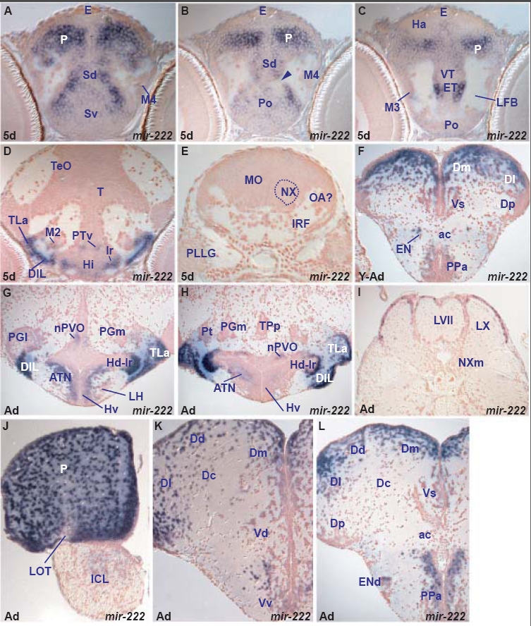
miR-222 expression is restricted to differentiating cells of the forebrain and midbrain and expression is largely conserved in these areas throughout life. In addition, miR-222 is de novo expressed in the adult facial and vagal lobes. In the larval brain, miR-222 is expressed in cells of all telencephalic areas apart from the lateral telencephalic area M4. In the diencephalon, miR-222 is expressed in the preoptic area, ventral thalamus and eminentia thalami, rostral and intermediate hypothalamic nuclei. More caudally, it is expressed in ventral and migrated posterior tubercular cells and a few scattered cells of the tectal periventricular gray zone (Table D). In the adult brain, miR-222 expression is conserved in the above areas with the exception of the thalamus where it is downregulated. In addition, miR-222 is expressed in the adult facial and vagal lobes although in the larva we did not observe expression in any hindbrain cells that could correspond to these adult areas (Table I).
A. transverse section through the larval telencephalon showing miR-222 expressing cells in the ventral (Sv) and dorsal (Sd) subpallium and pallium (P).
B. transverse section through the larval telencephalon (caudal to section A, at the level of the anterior commissure, arrowhead) showing miR-222 expressing cells in the preoptic area (Po), dorsal subpallium (Sd) and pallium (P).
C. transverse section through the larval caudal telencephalon and rostral diencephalon showing miR-222 expressing cells in the preoptic area (Po), eminentia thalami (ET), ventral thalamus (VT) and pallium (P).
D. transverse section through the larval hypothalamus and midbrain showing miR-222 expressing cells in the intermediate hypothalamus (Hi, ventral and at the level of lateral ventricular recess-lr), diffuse nucleus of the hypothalamic inferior lobe (DIL), lateral torus (TLa), ventral periventricular (PTv) and migrated posterior tubercular area (M2).
E. transverse section through the larval hindbrain at the level of the posterior lateral line ganglion (PLLG) where the medulla oblongata (MO) is devoid of miR-222 expression. The dorsal medulla oblongata at this level may give rise to cells of the facial and vagal lobes.
F. transverse section through the young adult caudal telencephalon at the level of the anterior commissure (ac) showing miR-222 expressing cells in the anterior parvocellular preoptic nucleus (PPa), endopeduncular nucleus (EN, part of eminentia thalami), supracommissural nucleus of the ventral telencephalon (Vs), medial (Dm) and lateral (Dl) zones of the dorsal telencephalon.
G. transverse section through the adult hypothalamus showing miR-222 expressing cells in the ventral zone of the periventricular hypothalamus (Hv), anterior tuberal nucleus (ATN), dorsal zone of the periventricular hypothalamus at the level of the lateral hypothalamic ventricular recess (Hd-lr), diffuse nucleus of the inferior hypothalamic lobe (DIL), lateral hypothalamic torus (TLa) and nucleus of the paraventricular organ (nPVO).
H. transverse section through the adult hypothalamus showing miR-222 expressing cells in the anterior tuberal nucleus (ATN), dorsal zone of the periventricular hypothalamus at the level of the lateral hypothalamic ventricular recess (Hd-lr), diffuse nucleus of the inferior hypothalamic lobe (DIL), lateral hypothalamic torus (TLa), nucleus of the paraventricular organ (nPVO) and posterior thalamic nucleus (Pt).
I. transverse section through the adult caudal hindbrain showing miR-222 expressing cells superficially lining the facial (LVII) and vagal (LX) lobes.
J. transverse section through the adult rostral telencephalon showing miR-222 expressing cells in the dorsal telencephalon/pallium (P) and internal cellular layer of olfactory bulb (ICL).
K. transverse section through the adult telencephalon showing miR-222 expressing cells in the ventral (Vv) and dorsal (Vd) nuclei of the ventral telencephalon (subpallium), medial (Dm), central (Dc), dorsal (Dd), lateral (Dl) and posterior (Dp) zones of the dorsal telencephalon.
L. transverse section through the adult telencephalon at the level of anterior commissure (ac) showing miR-222 expressing cells in the anterior parvocellular preoptic nucleus (PPa), dorsal part of the entopeduncular nucleus (ENd, part of the eminentia thalami), supracommissural/posterior nucleus of the ventral telencephalon (Vs), medial (Dm), dorsal (Dd) and lateral (Dl) zones of the dorsal telencephalon.

