
|
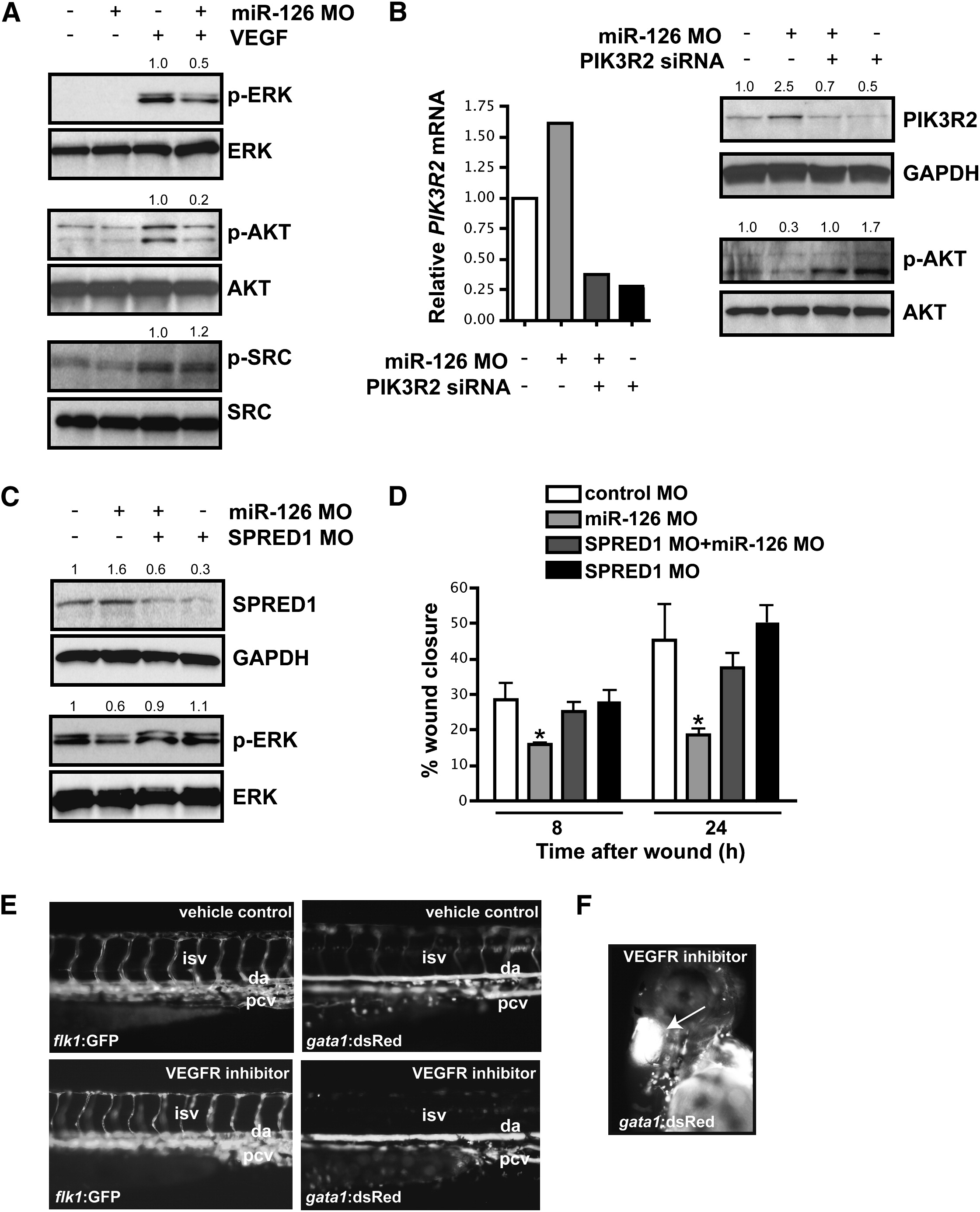
Fig. 5 miR-126 Positively Regulates VEGF Signaling in Endothelial Cells by Repressing SPRED1 and PIK3R2
(A) Immunoblot of lysates from HUVECs transfected with control or miR-126 MOs in the presence or absence of VEGF (10 ng/ml, 10 min). VEGF induced phosphorylation of ERK (p-ERK) and AKT (p-AKT), which was blocked by miR-126 inhibition. Total levels of ERK and AKT were not affected. Phosphorylation of SRC was not affected by miR-126 knockdown. Densitometric analysis of normalized protein levels are indicated above immunoblot.
(B) PIK3R2 mRNA was knocked-down by RNAi in HUVECs transfected with control or miR-126 MOs (qRT-PCR). Western analysis indicates a decrease in PIK3R2 protein by introduction of siRNA, even in the presence of miR-126 MOs. Knockdown of PIK3R2 rescued the defect in VEGF-dependent phosphorylation of AKT in miR-126 MO-treated cells.
(C) Western analysis shows reduced SPRED1 levels by transfection of a MO that blocks SPRED1 translation, even in the presence of miR-126 MO. SPRED1 MO rescued the defect in VEGF-induced phorphorylation of ERK in miR-126 MO-transfected cells.
(D) Quantification of percent (%) wound closure of endothelial cells in a ?scratch? assay reveals rescue of miR-126 MO effects by knocking down SPRED1. * p < 0.05 compared to control MO.
(E) Treatment of 48 hpf embryos with a VEGF receptor inhibitor (Vatalanib, 5 μM, 18 hr) resulted in reduced circulation of blood cells (gata1:dsRed). ISVs (flk1:GFP) appeared to lack a lumen after VEGF inhibition.
(F) Hemorrhages also occurred in some embryos treated with VEGF receptor inhibitor (arrow).
Reprinted from Developmental Cell, 15(2), Fish, J.E., Santoro, M.M., Morton, S.U., Yu, S., Yeh, R.F., Wythe, J.D., Ivey, K.N., Bruneau, B.G., Stainier, D.Y., and Srivastava, D., miR-126 regulates angiogenic signaling and vascular integrity, 272-284, Copyright (2008) with permission from Elsevier. Full text @ Dev. Cell