
|
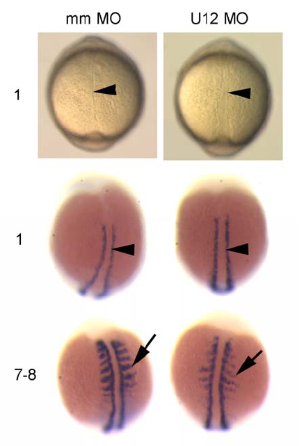
Fig. S5
Developmental Patterning of the Midline and Somite Segmentation Are Unaffected in U12-MO-Injected Embryos during Early Somitogenesis
Top: Bright field view from dorsal onto 1 somite stage embryos. Arrowhead indicates the notochord. Middle: myoD expression in one somite stage embryos shown from dorsal view. Arrowhead indicates the notochord, purple staining indicates the paraxial mesoderm. No difference is seen between mismatch- (mm) and U12-morpholino (MO) injected embryos in the formation of the notochord or in myoD expression during the start of somitogenesis. Bottom: myoD expression in 7-8 somite stage embryos shown from dorsal view. Arrow indicates somite boundaries, which are formed but with reduced myoD staining in U12 MO injected embryos. Numbers on the left indicate somite numbers of the embryos shown.
Reprinted from Cell, 131(4), K—nig, H., Matter, N., Bader, R., Thiele, W., and M■ller, F., Splicing Segregation: The Minor Spliceosome Acts outside the Nucleus and Controls Cell Proliferation, 718-729, Copyright (2007) with permission from Elsevier. Full text @ Cell