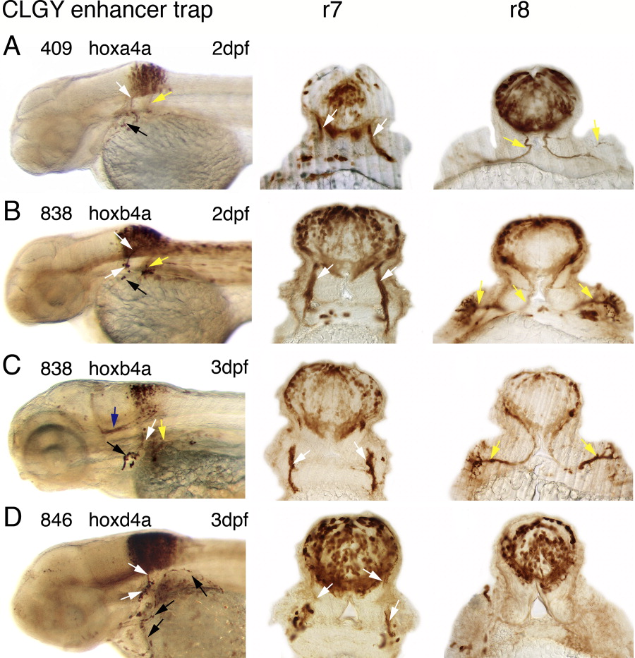
Fig. 3 Immunostained embryos/larvae of hoxa4a (A), hoxb4a (B, C), and hoxd4a (D) enhancer detection lines with transverse sections on the level of the first (r7) and third somite (r8). A: hoxa4a-regulated YFP expression at 2 dpf in the hindbrain r7 and r8, in the vagal nerve (white arrows), the pectoral fin nerve (yellow arrows), and in some neural crest cells (black arrow). Stained cells are located further ventrally and medially than in the hoxb4a enhancer trap line. B: hoxb4a-YFP expression in embryos at 2 dpf in the posterior hindbrain, in the vagal nerve, vagal ganglion (white arrows), in the pectoral fin nerve (yellow arrows), and some neural crest cells (black arrow). In r8, at the level of the fin motoneuron, there were no YFP-positive neurons in the medial hindbrain, which was also observed at later developmental stages. C: At 3 dpf, hoxb4a-regulated YFP was detected in ventrally located fiber tracts that grew from the posterior hindbrain towards the cerebellum (blue arrow). The white arrows point to the vagal and the yellow arrows to the pectoral fin nerve. Neural crest are marked by the black arrow. D: Hoxd4a-YFP expression at 3 dpf in the posterior hindbrain, in the vagal nerve and the vagal ganglion (white arrows), in the heart wall and the fin (black arrows). In contrast to the hoxb4a enhancer detection line, cells are also stained in the medioventral hindbrain.
Image
Figure Caption
Figure Data
Acknowledgments
This image is the copyrighted work of the attributed author or publisher, and
ZFIN has permission only to display this image to its users.
Additional permissions should be obtained from the applicable author or publisher of the image.
Full text @ Dev. Dyn.

