
Image
|
Figure Caption
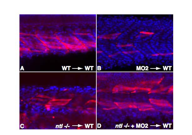
Fig. S3
Muscle Morphology of Transplanted Cells
High magnification images of embryos shown in Figure 3 reveal elongated myotube morphology of transplanted cells (red) in tail somites. Nuclei are stained with DAPI (blue). All embryos are shown from a lateral view with anterior to the left.
Acknowledgments
This image is the copyrighted work of the attributed author or publisher, and
ZFIN has permission only to display this image to its users.
Additional permissions should be obtained from the applicable author or publisher of the image.
Reprinted from Developmental Cell, 15(1), Martin, B.L., and Kimelman, D., Regulation of canonical Wnt signaling by Brachyury is essential for posterior mesoderm formation, 121-133, Copyright (2008) with permission from Elsevier. Full text @ Dev. Cell