
|
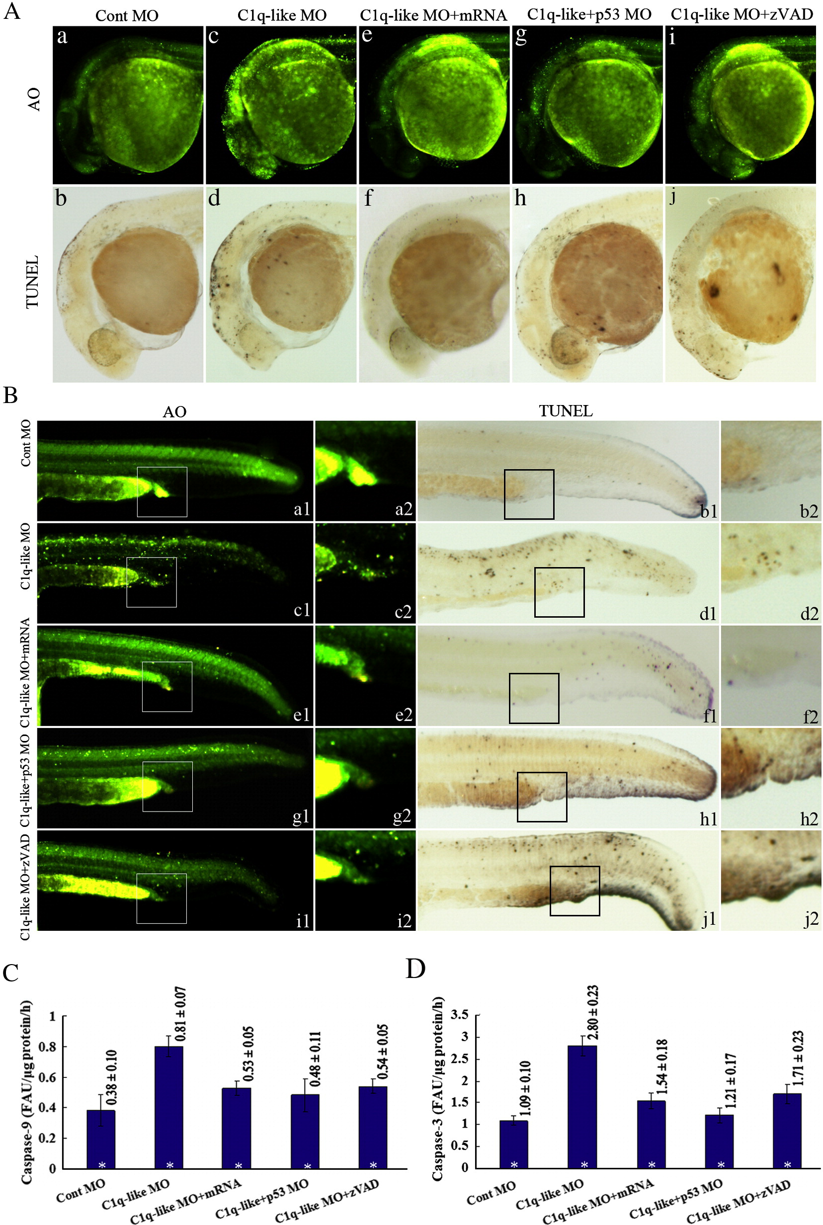
Fig. 4 Developmental defects and caspase 3/9 activity changes in the C1q-like MO embryos. (A) The head phenotypes revealed by AO staining (a, c, e, g, i) and TUNEL assays (b, d, f, h, j). The pictures are representative of at least three experiments (more than 30 embryos in each experiment). (B) The tail phenotypes revealed by AO staining (a, c, e, g, i) and TUNEL assays (b, d, f, h, j). The picture a2?j2 showing the corresponding amplification signals of the apoptotic cells in intermediate cell mass (ICM) region from a1?j1. (C, D) Caspase-9 (C) and 3 (D) activity changes in the shown experiments. The activities were measured from embryos at 28 hpf by cleavage of AFC from synthetic substrate, were expressed as change in FAU/μg protein/h. The data represent mean ± SD of three separate experiments from thirty injected embryos. *p < 0.05.
Reprinted from Developmental Biology, 319(2), Mei, J., Zhang, Q.Y., Li, Z., Lin, S., and Gui, J.F., C1q-like inhibits p53-mediated apoptosis and controls normal hematopoiesis during zebrafish embryogenesis, 273-284, Copyright (2008) with permission from Elsevier. Full text @ Dev. Biol.