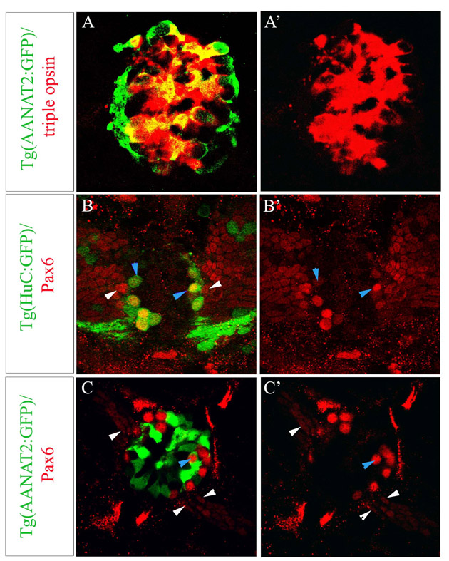
Fig. S1 (A-A′) Optical sections of Tg(AANAT2:GFP) epiphysis at 2 days of development stained with a triple red opsin, exorhodopsin, rhodopsin probe and GFP. All opsin-positive cells are Tg(AANAT2:GFP)+; conversely, only 87.2±4.14% of Tg(AANAT2:GFP)+ cells are positive for one of the three opsins. (B-C′) Optical sections of Tg(HuC:GFP) (B,B′) and Tg(AANAT2:GFP) (C,C′) epiphyses at 48 hours labelled with a Pax6 antibody and GFP. Tg(HuC:GFP)+ cells are also positive for Pax6 (blue arrowheads). Numerous Pax6+, Tg(HuC:GFP)- neuroepithelial cells are observed in the ventrolateral part of the epiphysial area (white arrowheads). Pax6 and Tg(AANAT2:GFP) expression is mostly exclusive except for rare cells expressing both markers (blue arrowheads).
Image
Figure Caption
Acknowledgments
This image is the copyrighted work of the attributed author or publisher, and
ZFIN has permission only to display this image to its users.
Additional permissions should be obtained from the applicable author or publisher of the image.
Full text @ Development

