
|
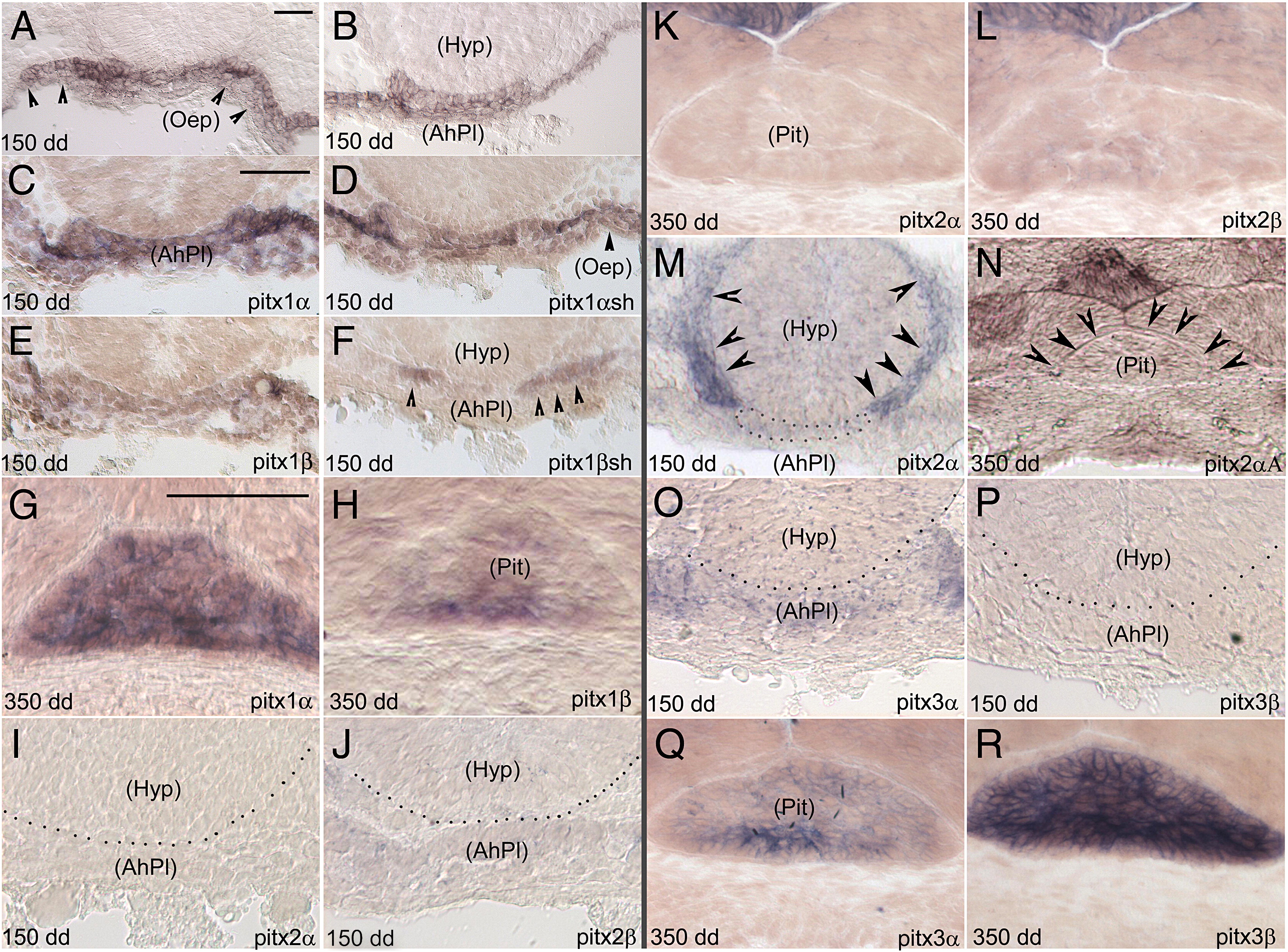
Fig. 4 Pituitary expression patterns of the salmon pitx- gene complement analyzed on serial sections at early and late stages of embryonic development. Gene names and stages of development are indicated on the lower-right and left corner of each panel, respectively. (A,B): pitx1 expression analysis using a probe cross-hybridizing with the four pitx1 transcripts. (C,D,E): pitx1α, pitx1αsh and pitx1β expression in the oral cavity epithelium (Oep), and in adenohypophyseal placode (AhPl). (F): pitx1βsh transcripts in the dorsal sides of the (AhPl). (I,J/O,P) and (K,L/Q,R): expression analysis of pitx2α/pitx2β and pitx3α/pitx3β paralog-pairs at 150 and 350 dd respectively. (M): note the circular expression domain of pitx2α in the hypothalamus (Hyp; marked by arrows), and the absence of expression in the adenohypophyseal placode (outlined by dots). (N): lack of pitx2αA gene expression in the developed pituitary; the border of the gland is indicated by arrows. Scale bar for (A,B), (C?F,M,N), (G?L) and (O?R) = 50 μm.
Reprinted from Gene, 417(1-2), Angotzi, A.R., Ersland, K.M., Mungpakdee, S., Stefansson, S., and Chourrout, D., Independent and dynamic reallocation of pitx gene expression during vertebrate evolution, with emphasis on fish pituitary development, 19-26, Copyright (2008) with permission from Elsevier. Full text @ Gene