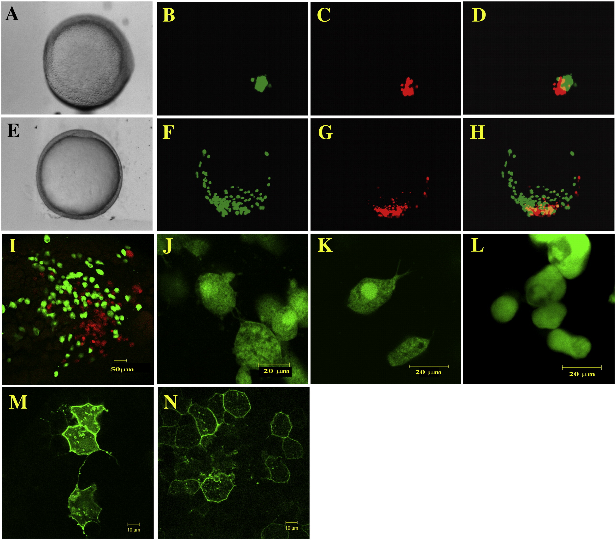
Fig. 8 Cell migration, behavior and shape are affected by Pcdh18a overexpression. Cells from embryos injected with 150–200 pg GFP RNA and 2.5 ng fluoro-emerald dextran (green cells), or 150–200 pg pcdh18a RNA and 5 ng fluoro-ruby dextran (red cells), were transplanted to the same region in the margin of uninjected host embryos at 40% epiboly (A–D), and cultured to 80–90% epiboly (E–H). Host embryos were examined in visible light (A, E) and by fluorescence microscopy (C, D, F, G; overlays in panels D, H). In 43% of the embryos examined, the green control cells migrated farther than the red Pcdh18a-expressing cells. (I) A different transplanted embryo was examined by confocal microscopy, showing the same effect at higher magnification (scale bar, 50 μm). (J–L) Confocal micrographs of cells from embryos injected with emerald dextran together with 150 pg of GFP RNA (J, K), or pcdh18a RNA (L), transplanted into separate host embryos. Cell protrusions are visible in control cells but are missing in the pcdh18a-expressing cells. Scale bar is 20 μm. Cells shown in panel J are from an embryo fixed and mounted in glycerol, while the cells shown in panels K and L are from live embryos mounted in low melt agarose. (M, N) 20 pg mGFP alone or 10 pg mGFP in combination with 10 pg pcdh18a RNAs was injected into one cell at the 64-cell stage. Confocal microscopy at 70% epiboly showed fewer protrusions in pcdh18a RNA-injected embryos compared to controls (M, N). Scale bar in panels M and N is 10 μm.
Reprinted from Developmental Biology, 318(2), Aamar, E., and Dawid, I.B., Protocadherin-18a has a role in cell adhesion, behavior and migration in zebrafish development, 335-346, Copyright (2008) with permission from Elsevier. Full text @ Dev. Biol.

