
|
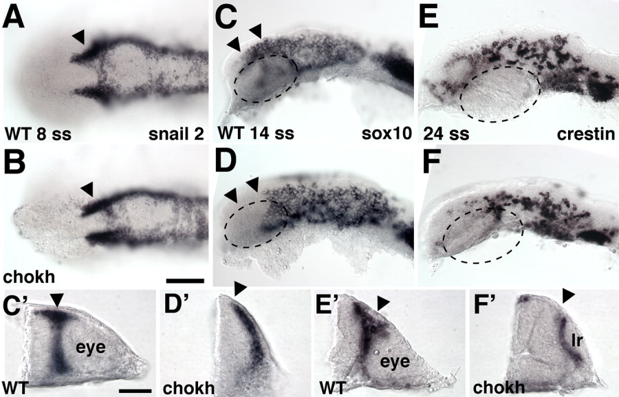
Fig. 4 Neural crest cells (NCCs) in chokh embryos. A-F: Wild-type (WT) and chokh mutant embryos labeled with in situ hybridization for NC markers at the indicated stages. Arrowheads mark normal (A-C,C′,E′) and missing (D,D′,F′) NCC populations. The hatched circle marks the eye in WT embryos (C,E) and the approximate position where the eye would normally have developed in chokh embryos (D,F). A, B are dorsal, C-F lateral views, anterior to the left. C′-F′: Cross-sections through the eye vesicle in WT and the respective region in chokh embryos. Scale bar = 100 μm in A-F, 50 μm in C′-F′. lr, lens remnant.