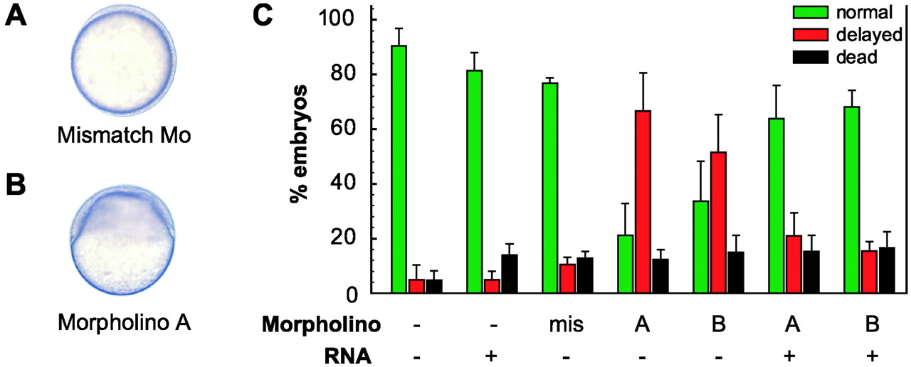
Fig. 5 Morpholino knockdown of zCOX-1. (A and B) Micrographs of zebrafish embryos (Normarski optics) at 11 hpf. Embryo (A) was injected with a control mismatch morpholino (0.85 ng) in the one-cell stage and has progressed normally to the tailbud stage. Embryonal cells have migrated around the yolk and the embryonal axis has been established. Embryo (B) was injected with 0.85 ng zCOX-1 antisense morpholino and is severely delayed in its development. Embryonal cells are covering only 40% of the yolk (40% epiboly). (C) Phenotypic rescue by zCOX-1 RNA. Embryos were classified as normally developed (green bars) or delayed (red bars) at 12 hpf. Lethality at 12 hpf is shown by the black bars. Embryos were microinjected in the one-cell stage with either mismatch morpholino (mis), 0.85 ng morpholino zCOX-1A (A), or 0.85 ng morpholino zCOX-1B (B). The ratio of delayed/normal embryos was reversed when COX-1 RNA (200 pg) was injected simultaneously with the antisense morpholinos as compared with the morpholinos alone. Each treatment group consists of injections of four different egg lays and minimally 160 individual embryos in total. Data are means (±SEM) of percent embryos in each set of delivered eggs.
Image
Figure Caption
Figure Data
Acknowledgments
This image is the copyrighted work of the attributed author or publisher, and
ZFIN has permission only to display this image to its users.
Additional permissions should be obtained from the applicable author or publisher of the image.
Full text @ Proc. Natl. Acad. Sci. USA

