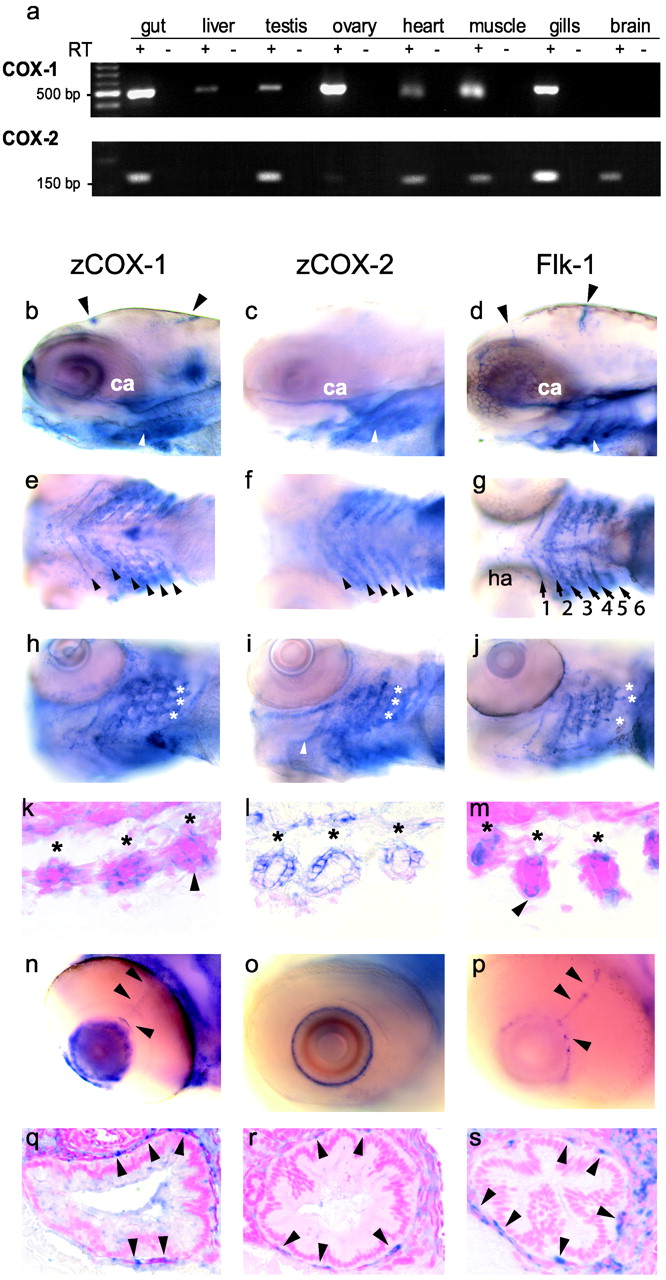
Fig. 4 (a) Analysis of zCOX-1 and zCOX-2 transcript expression in adult zebrafish. We assessed constitutive zCOX isoform expression in adult zebrafish by reverse transcriptase-PCR using total RNA from freshly dissected adult organs. PCR products (500 bp and 162 bp) and the corresponding negative controls were visualized by agarose gel-electrophoresis. Expression of zCOX-1 (b, e, h, k, n, and q), zCOX-2 (c, f, i, l, o, and r), and flk-1 (d, g, j, m, p, and s) by whole-mount in situ hybridization in zebrafish larvae at 96 hpf. zCOX-1 (b), zCOX-2 (c), and the endothelial marker flk-1 (d) are expressed in the carotid artery (ca) and the pharyngeal arches (white arrow) (lateral view). zCOX-1 (b) and flk-1 (d) are also present in cranial arteries (arrowheads). (e-g) Ventral views showing expression in the pharyngeal arches (arrowheads). While flk-1 staining (g) highlights the vasculature in the center of the arches [arch arteries 1 and 2 (opercular artery); 3-6,ha, hypobranchial artery (35)], zCOX-1 (e) and zCOX-2 (f) appear to be expressed in more peripheral structures of the arches. (h-j) Oblique ventral views. zCOX-1 (h), zCOX-2 (i), and flk-1 (j) colocalize and are expressed within sprouting gill arteries (*) that are apparent as protrusions at the caudal surface of arch arteries 3-6. (k-m) Histological cross sections through these vessels (*, section rotated by 90°) show flk-1 (m) expression in endothelial cells. zCOX-2 is highly expressed in the vessel wall of the sprouting gill vasculature (l). zCOX-1 expression is less intense and appears to be present in both endothelial and wall structures (k). (n-p) Endothelial expression of zCOX-1 was seen the ciliary arteries of the eye (n, arrowheads), which were also visible in flk-1 (p), but not in zCOX-2-stained embryos (o). zCOX-1 and zCOX-2 were both differentially expressed in other structures of the developing eye (n and o). (q-s) All three genes were expressed in the vasculature of the intestine. Endothelial cells are highlighted by arrowheads. (Magnifications: (b-j) x200, (k-m and q-s) x630, n-p) x400.)
Image
Figure Caption
Figure Data
Acknowledgments
This image is the copyrighted work of the attributed author or publisher, and
ZFIN has permission only to display this image to its users.
Additional permissions should be obtained from the applicable author or publisher of the image.
Full text @ Proc. Natl. Acad. Sci. USA

