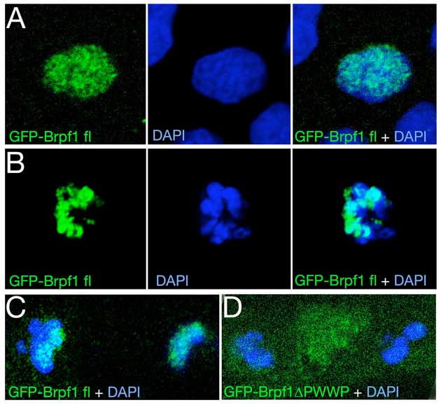
Fig. S7 Brpf1-GFP displays PWWP domain-dependent chromatin association in zebrafish embryos. Zebrafish embryos were injected at the 1-cell stage with mouse full-length GFP-Brpf1 or GFP-Brpf1ΔPWWP plasmids, as also used for Fig. S1E, Figs 5 and 6, and fixed at mid-gastrula stages (7-9 hpf). GFP was visualized by immunofluorescence, as described for transfected HEK 293 cells (Figs 5 and 6), DNA was counterstained with DAPI, and embryos were analyzed by confocal microscopy (Zeiss LSM510 META). (A-C) Full-length Brpf1; (D) Brpf1ΔPWWP. (A) Interphase, (B) early mitosis, (C,D) late mitosis, with the two sets of sister chromosomes clearly separated. Full-length Brpf1 is localized at discrete sites of the chromatin of zebrafish cells both in interphase (A) and mitosis (B,C); compare with Fig. 5A and Fig. 6B for HEK 293 cells. (C,D) In contrast to full-length Brpf1 (C), Brpf1ΔPWWP lacking the PWWP domain is largely excluded from the chromatin (D); compare with Fig. 6D.
Image
Figure Caption
Acknowledgments
This image is the copyrighted work of the attributed author or publisher, and
ZFIN has permission only to display this image to its users.
Additional permissions should be obtained from the applicable author or publisher of the image.
Full text @ Development

