
Fig. 3
|
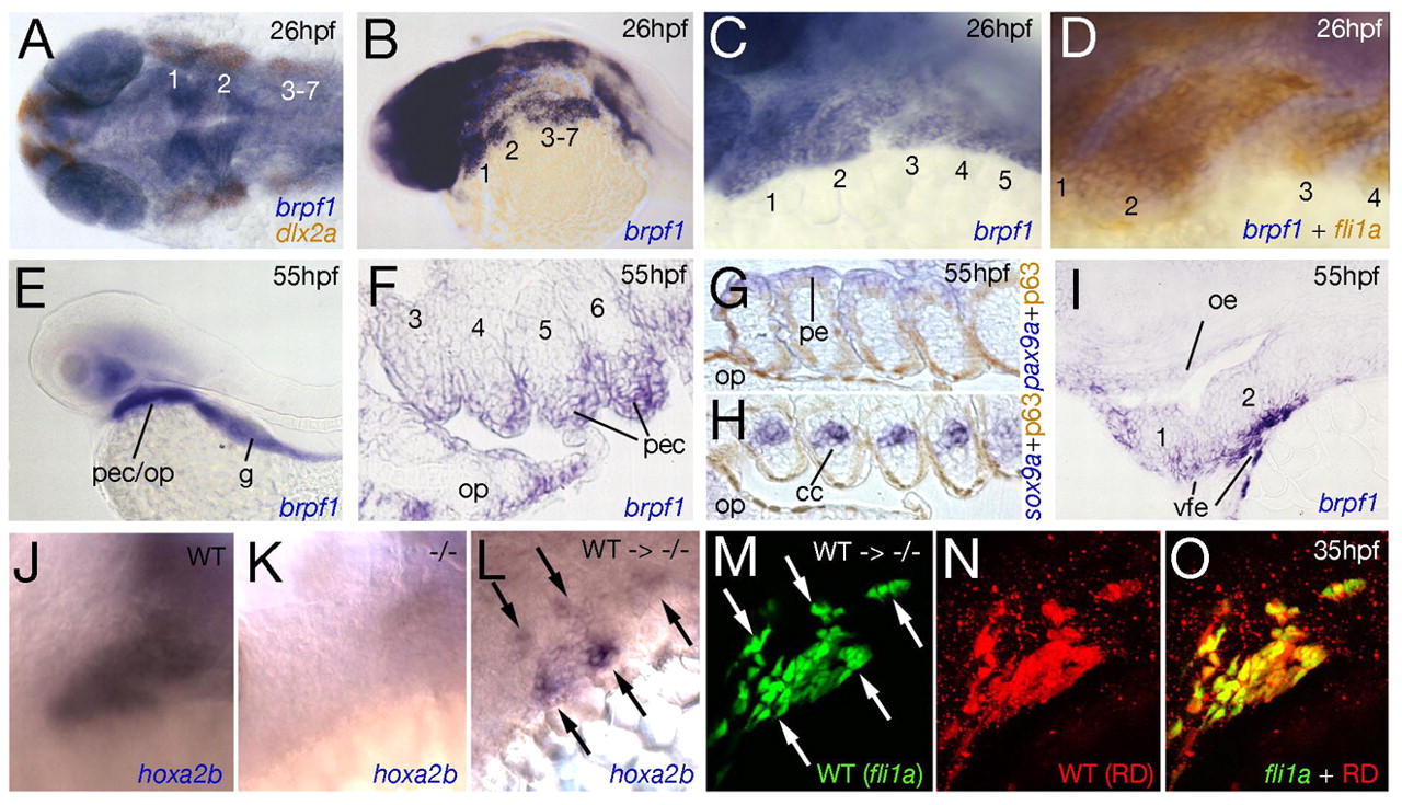
Fig. 3 Expression pattern and cell-autonomous function of brpf1 in zebrafish CNC. Staining with reagents indicated at lower right at the stages indicated upper right. Numbers of pharyngeal arches are indicated (1-7). (A-J) Wild-type embryos; (K) brpf1b943 mutant (-/-); (L-O) mutant transplanted with wild-type cells (WT → -/-). (A) Dorsal view; (B-E,J-O) lateral views; (F-H) horizontal section; (I) longitudinal section. (A-D) At 26 hpf, brpf1 is co-expressed with the CNC marker dlx2a (A) and with fli1a (D), stained by anti-GFP immunostaining of tg(fli1a:EGFP) embryo (Isogai et al., 2003). brpf1-positive cells between CNC include pharyngeal endoderm [D; compare with Fig. 1A in Crump et al. (Crump et al., 2004)]. (E-I) At 55 hpf, sox9a-positive chondrocytes of cartilage condensates (cc; H) (Yan et al., 2002) and pax9a-positive pharyngeal endodermal cells (pe; G) (Nornes et al., 1996; Okabe and Graham, 2004) lack brpf1 expression, which, however, is strongly expressed in p63 (tp63 - ZFIN)-positive cells (Carney et al., 2007) of the pharyngeal ectoderm (pec; F-H), in the oral ectoderm (oe; I) and in facial ectoderm ventral to arches 1 and 2 (vfe; I) (Crump et al., 2006). (J-O) Analysis of chimeric embryos with rhodamine-dextran (RD)-labeled (N) and tg(fli1a:EGFP)-positive (M) wild-type cells integrated in the CNC of a brpf1 mutant host [for procedure, see Crump et al. (Crump et al., 2006)]. Only wild-type, not adjacent mutant CNC, cells display hoxa2b expression (arrows in L and M; n=3/3). g, gut; op, opercle.