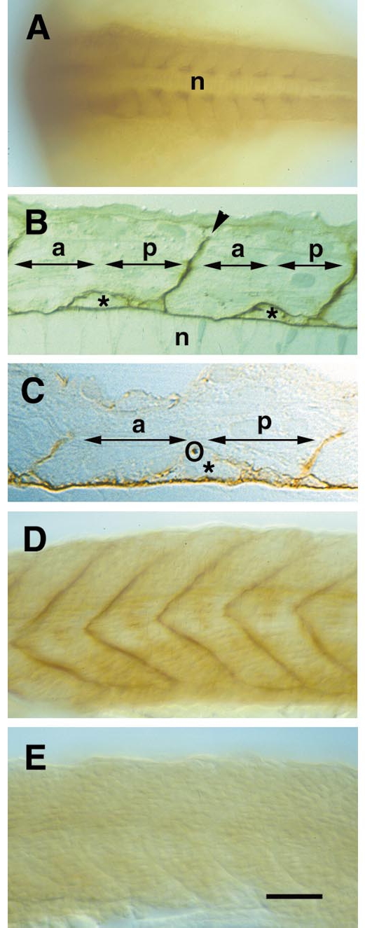
Fig. 2 The distribution of chondroitin sulfate immunoreactivity in the zebrafish embryo and its elimination by chondroitinase treatment are shown by immunostaining using mAb 473. (A, D, and E) Whole-mount preparations; (B and C) semithin sections. Anterior is to the left, in D and E dorsal is to the top. (A) A low-power dorsal view of an 18-hpf embryo shows accumulation of chondroitin sulfate labeling in the posterior half of each somite, adjacent to the notochord (n). The segmental myosepta are also labeled. (B) A horizontal section through a 22-hpf embryo reveals chondroitin sulfate reactivity around the notochord (asterisk), around putative migrating sclerotome cells (arrows), and in the segmental myosepta (arrowhead). (C) A high-power view of a horizontal section through a 24-hpf embryo shows the relationship of the ventral motor nerve (labeled by the tubulin antibody, circled) to putative migrating cells and chondroitin sulfate immunoreactivity which appears relatively faint in this doubly labeled preparation. A comparison with B will confirm, however, that the migrating cells along which the axons grow are surrounded by chondroitin sulfates. (D) A side view of a 26-hpf embryo shows the prominent chondroitin sulfate reactivity in the segmental myosepta. (E) Similar view of a corresponding embryo injected with chondroitinase ABC at 16 hpf. No chondroitin sulfate reactivity can be detected. Magnification bar: 100 μm for (A), 25 μm for (B), 10 μm for (C), and 70 μm for (D and E).
Reprinted from Developmental Biology, 221(1), Bernhardt, R.R. and Schachner, M., Chondroitin sulfates affect the formation of the segmental motor nerves in zebrafish embryos, 206-219, Copyright (2000) with permission from Elsevier. Full text @ Dev. Biol.

