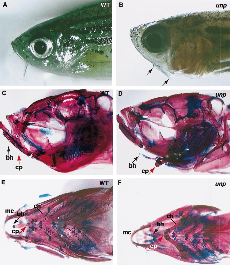
Fig. 7 Pharyngeal arch defects in unplugged mutant adult fish. Lateral views of 17-week-old wild-type (A) and unplugged mutant (B) fish. In unplugged mutant fish (B), a ventral protrusion is visible from the lower jaw (black arrows in B). Skeletal preparations of the same wild-type (C, E) and unplugged mutant (D, F) adult fish. Lateral (C, D) and ventral (E, F) views of wild-type (C, E) and unplugged fish stained for chondrocytes (blue) and calcified bones (red). (C) In wild-type fish, the basihyal (bh; black arrow) and its connection point (cp; red arrow) with the ceratohyals (ch) are not distinguishable in a lateral view, and only their approximate locations are indicated. (D) In unplugged mutant fish, the basihyal (black arrow) and its connection point with the ceratohyals (red arrow) are located more ventrally and posteriorly. (E) Ventral view of the same wild-type preparation shown in C. (F) Ventral view of the same unplugged preparation shown in D. Note the increased distance between Meckel’s cartilages (mc) and the anterior tip of the basihyal (bh, black arrow), compared to wild-type fish (E). cp, connection point between the ceratohyals and the basihyal; bh, basihyal; ch, ceratohyal; mc, Meckel’s cartilages.
Reprinted from Developmental Biology, 240(2), Zhang, J., Malayaman, S., Davis, C., and Granato, M., A dual role for the zebrafish unplugged gene in motor axon pathfinding and pharyngeal development, 560-573, Copyright (2001) with permission from Elsevier. Full text @ Dev. Biol.

