
|
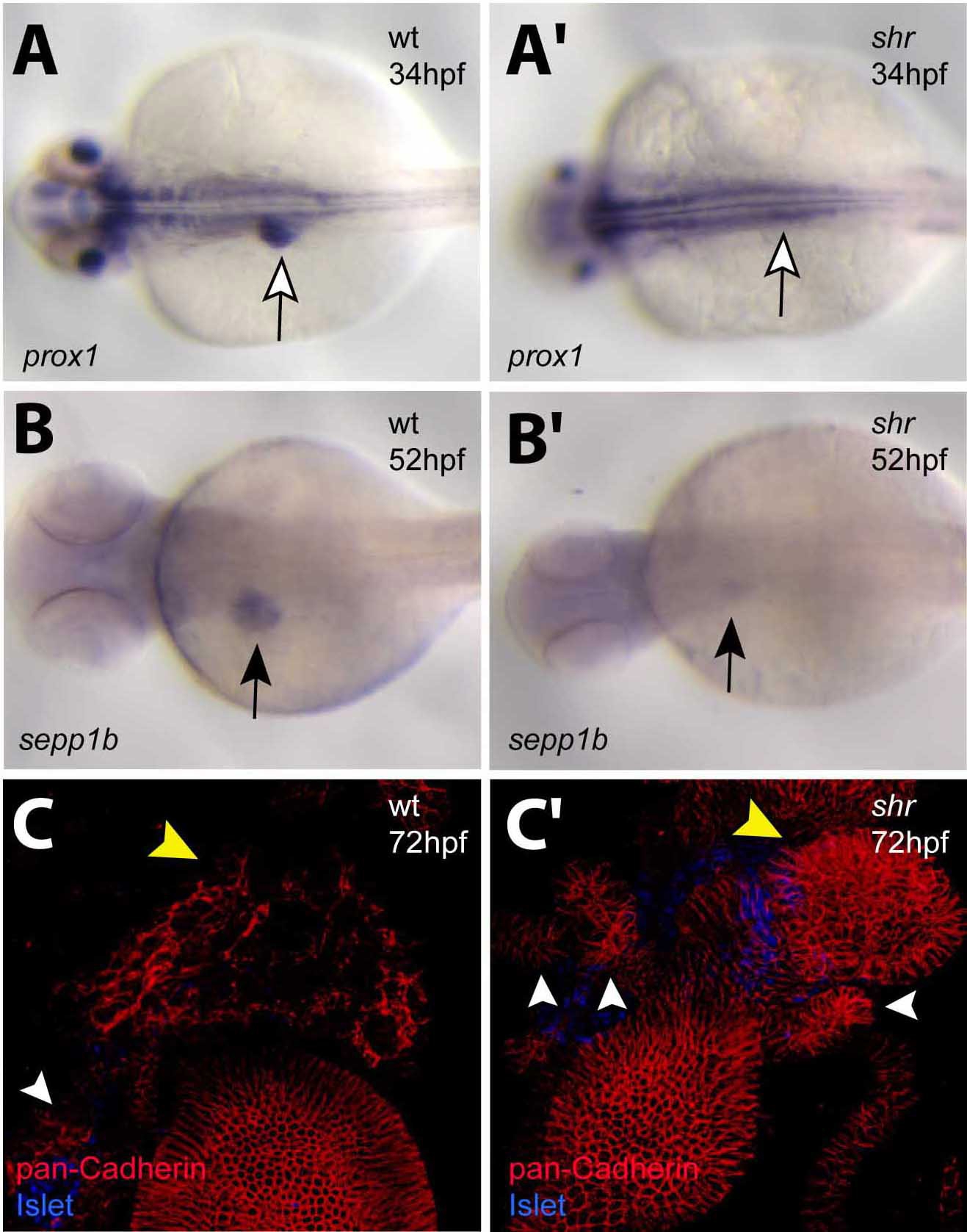
Fig. S1 shr is required for liver development. Wild-type and shrs435 mutant animals at 34 (A, A′), 52 (B, B′), and 72 (C, C′) hpf were analyzed for hhex (A, A′) and sepp1b (B, B′) expression and confocal projections of pan-Cadherin (red) and Islet (blue) expression (C, C′). (A, A′) prox1 expression is evident in the liver (white arrow) of wild-type embryos but barely detectable in the liver region of shrs435 mutants. (B, B′) selenoprotein P, plasma, 1b (sepp1b) expression is detected at low levels in shrs435 mutants (black arrow). (C, C′) shrs435 mutants exhibit defective differentiation of hepatocytes as assessed by Cadherin immunostaining. Note the multiple, unfused ventral pancreatic buds in mutants (white arrowheads). Yellow arrow points to the liver. A?B′ are dorsal views, anterior to the left; C, C′ are ventral views, anterior to the top.
Reprinted from Developmental Biology, 317(2), Shin, C.H., Chung, W.S., Hong, S.K., Ober, E.A., Verkade, H., Field, H.A., Huisken, J., and Stainier, D.Y., Multiple roles for Med12 in vertebrate endoderm development, 467-479, Copyright (2008) with permission from Elsevier. Full text @ Dev. Biol.