
|
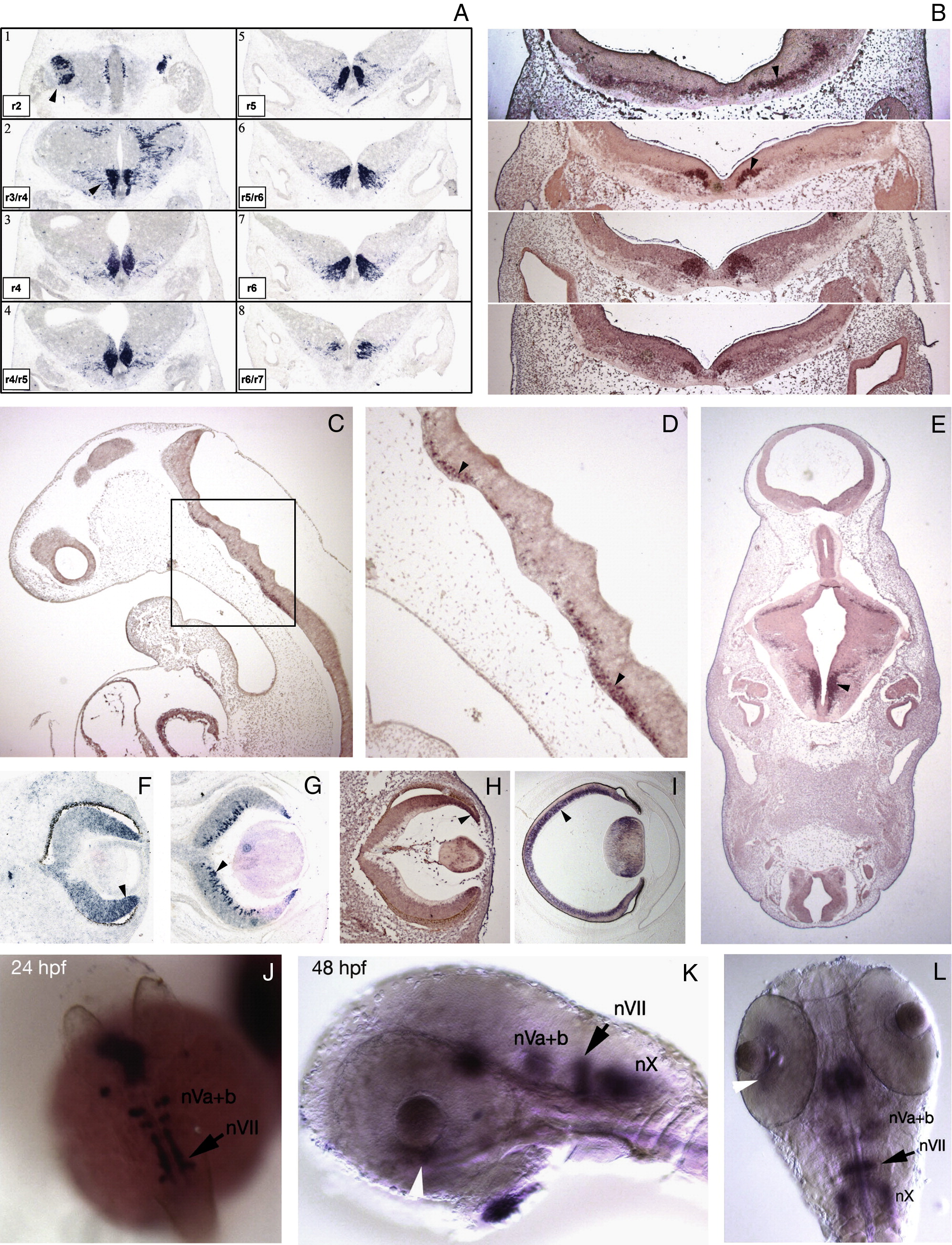
Fig. 2 Expression of vertebrate Tbx20 orthologues. (A) Tbx20 expression in E11.5 mouse hindbrain in dorsolaterally migrating trigeminal motor neurons (plate 1; arrowhead) and caudo-laterally migrating facial motor neurons (plates 2–8; arrowhead) in transverse sections at the level of rhombomeres (r) 2 to 7. (B–E) TBX20 expression in migrating motor neurons in the hindbrain of human embryos (arrowheads). (B) Series of rostral to caudal transverse sections through the hindbrain at 120-μm intervals, CS15, 33 days. (C, D) CS13, 28 days. (E) CS16, 37 days. Panel D shows high magnification view of boxed area in panel C. (F–I) TBX20/Tbx20 expression in mouse and human neural retina (arrowheads). (F) E11.5 (mouse). (G) E15.5 (mouse). (H) CS16, 37 days (human). (I) fetal stage 2, 63 days (human). (J–L) tbx20 expression in cranial motor neurons in zebrafish embryos at 24 (J) and 48 hpf (K, L). Black arrows indicate facial motor neurons (nVII). Trigeminal motor neurons (nVa + b) and vagal motor neurons (nX) are also shown. White arrowheads in panels K and L indicate retinal neurons.
Reprinted from Developmental Biology, 317(2), Pocock, R., Mione, M., Hussain, S., Maxwell, S., Pontecorvi, M., Aslam, S., Gerrelli, D., Sowden, J.C., and Woollard, A., Neuronal function of Tbx20 conserved from nematodes to vertebrates, 671-685, Copyright (2008) with permission from Elsevier. Full text @ Dev. Biol.