
|
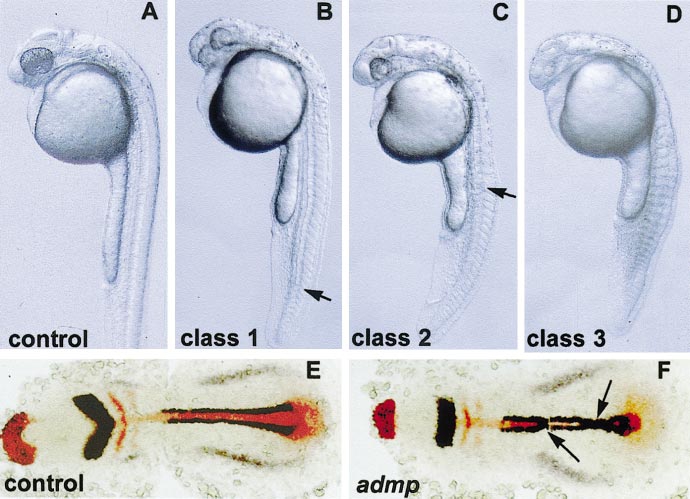
Fig. 3 admp overexpression causes the loss of dorsal fates. wt embryos were injected at the one-cell stage with admp RNA (600 pg) and compared to uninjected siblings (control). (A?D) Live embryos at 1 day of development. (E, F) Flat mounts of embryos stained at the one-to three-somite stage with myoD and pax2.1 in dark blue, hgg1, krox20, and ntl in red; anterior to the left. (A) Uninjected embryo. (B?D) admp-injected embryos. (B) Class 1 phenotype. (C) Class 2 phenotype. (D) Class 3 phenotype. (B, C) Arrows indicate the posterior limit of the truncated notochord. (E) Uninjected embryo. (F) admp-injected embryo; notochord defects and fusion of myoD expression domains at the midline are indicated (arrows).
Reprinted from Developmental Biology, 241(1), Willot, V., Mathieu, J., Lu, Y., Schmid, B., Sidi, S., Yan, Y.-L., Postlethwait, J.H., Mullins, M., Rosa, F., and Peyri�ras, N., Cooperative action of ADMP- and BMP-mediated pathways in regulating cell fates in the zebrafish gastrula, 59-78, Copyright (2002) with permission from Elsevier. Full text @ Dev. Biol.