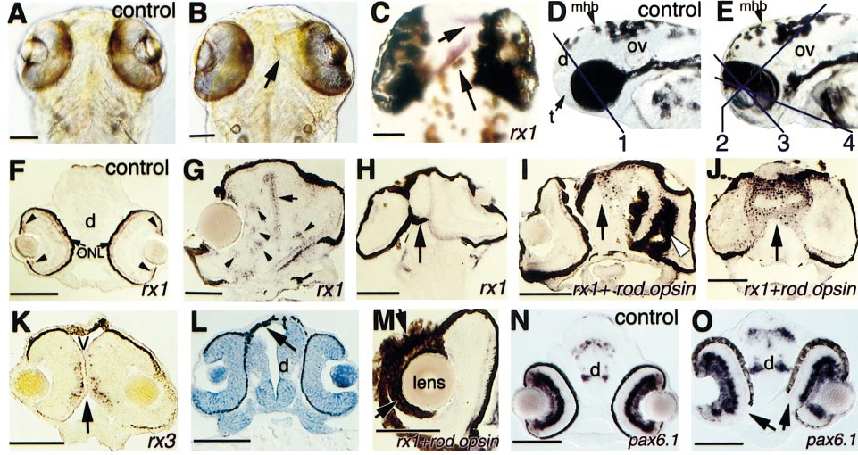
Fig. 6 Abnormalities of eye development following rx2 overexpression. (A, B) Comparison of eye morphology in control and rx2-injected embryos (PTU-treated, dorsal view) at 88 hpf. The arrow in (B) indicates medial expansion of the right eye. (C) Ectopic expression of rx1 (arrowheads) in the dorsal forebrain. (D, E) Comparison of control and rx2-injected embryos (lateral view). Lines in (D) and (E) represent the estimated section planes shown in (F, N): 1; (H): 2; (G, I?L, O): 3; and (M): 4. (F) In control embryos at 74 hpf, rx1 is expressed in the outer nuclear layer and circumferential germinal zone (arrowheads) of the neural retina. (G) Ectopic cone photoreceptors (expressing rx1) form rosettes within neural retina (arrowheads) and are found at the boundary between the fused eyes (arrow). (H) The entire rostral end of this embryo (arrow) consists of retinal tissue. (I) Ectopic RPE and photoreceptors (arrow) are found in forebrain, and ectopic RPE is in neural retina (arrowhead). (J) Ectopic photoreceptors (arrow) are in forebrain. (K) No forebrain tissue is found between the two eyes (arrow). In the differentiated retina, rx3 is expressed in the inner nuclear layer (Chuang et al., 1999). (L) Replacement of telencephalic tissue by ectopic RPE (arrow; methylene blue staining). (M) Ectopic RPE is surrounding the lens. (N?O) Comparison of pax6.1 expression patterns between control and injected embryos. The arrows in (O) show the lack of RPE and expansion of neural retina ventrally. Embryos were injected with 300 pg rx2ΔRx (B, E), 600 pg myc-rx2 (C, G?J, L, M), or 50 pg rx2ΔN′ (K, O) RNA, and examined at 88 hpf (A, B), 78 hpf (C, G, J), 60 hpf (D, E, N, O), 74 hpf (F, K), 96 hpf (H, I, M), and 46 hpf (L). d, diencephalon; mhb, midbrain hindbrain boundary; ONL, outer nuclear layer; ov, otic vesicle; t, telencephalon; V, ventricle. Scale bars: 100 μm.
Reprinted from Developmental Biology, 231, Chuang, J.-C. and Raymond, P.A., Zebrafish genes rx1 and rx2 help define the region of forebrain that gives rise to retina, 13-30, Copyright (2001) with permission from Elsevier. Full text @ Dev. Biol.

