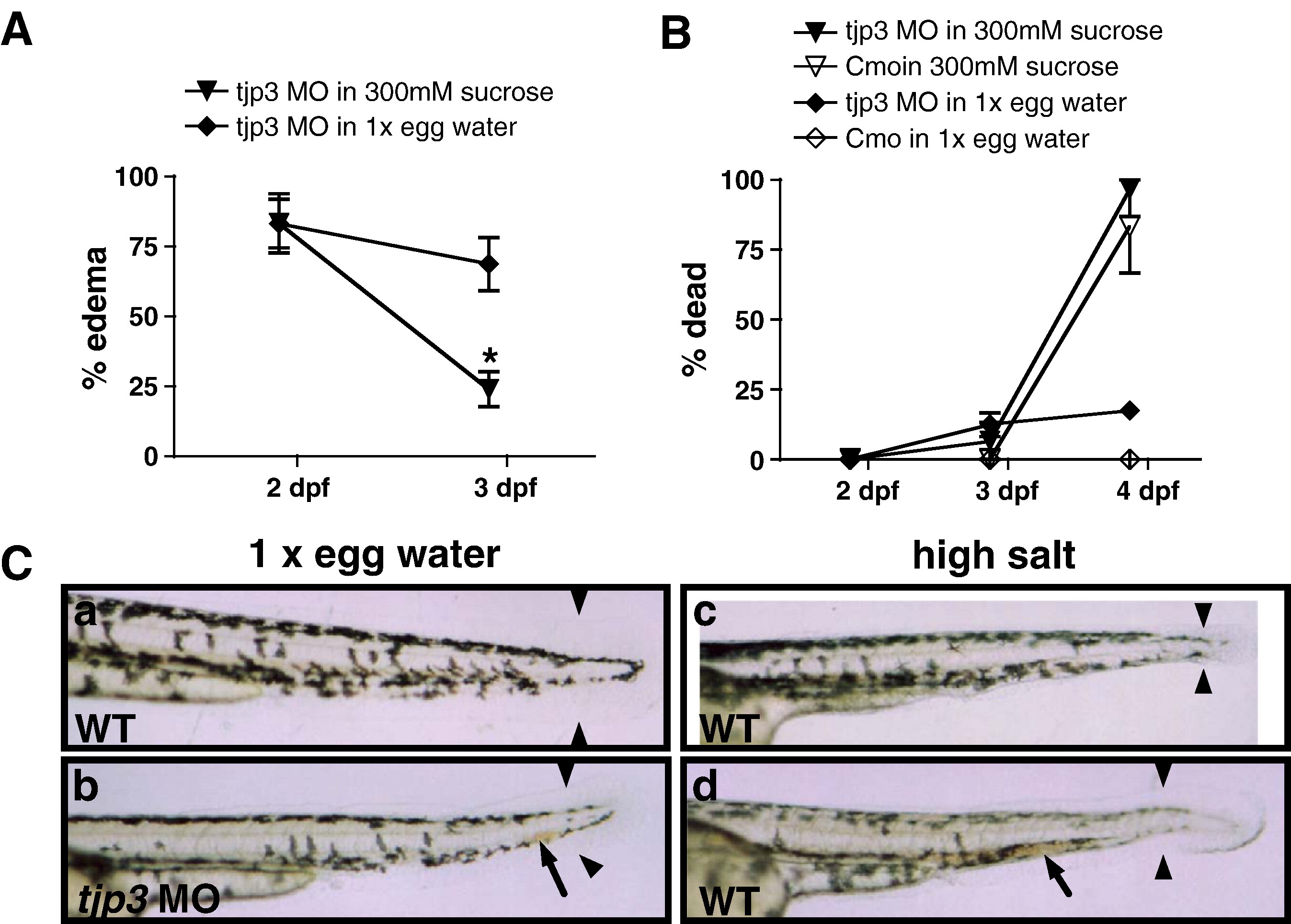
Fig. 10 Compromised osmotic homeostasis mimics the tjp3/zo-3 morphant phenotype. (A) Pericardial edema of tjp3/zo-3 morphants can be reversed by growing the embryos in a hyperosmotic sucrose solution. At 2 dpf tjp3/zo-3 morphants were transferred into a 300 mM sucrose solution. ∼ 80% of tjp3/zo-3 morphants presented with edema (∼ 20% had a curved tail only). If grown in 1x egg water (diamonds), the incidence of edema only slightly decreased, due to individuals that died. However, if tjp3/zo-3 morphants were grown in 300 mM Sucrose (triangles), edema incidence dropped significantly. Mean and S.E.M.; n ≥ 10 per experiment; 5?6 independent experiments. 2-way ANOVA; p < 0.01. (B) Mortality of embryos grown in 300 mM sucrose. Morpholino injected embryos grown in 1x egg water had a mortality of only ∼ 20% at 4 dpf (diamonds), whereas the mortality for controls was very low (open diamonds). Placed in a hyperosmotic sucrose solution at 2 dpf, most morpholino injected (triangles) and control (open triangles) embryos survived up to 3 dpf, when edema incidence was counted. However, after 48 h exposure close to 90% of both control and morphant embryos died. There was no statistical significance between mortality of control or morphant embryos in 300 mM sucrose. Mean and S.E.M.; n ≥ 10 per experiment; 2?6 independent experiments. Two-way ANOVA; p > 0.5. (C) Exposure of WT embryos to high salt conditions mimics the tjp3/zo-3 morphant phenotype. WT embryos in 1x egg water have a straight tail, a wide tailfin and circulating blood (a). Tjp3/zo-3 morphants in 1x egg water presented with curved tail, reduced tailfin (b, arrowheads), the loss of blood circulation and an accumulation of red blood cells (b, arrow). 10?15% of control embryos raised in 100x egg water (c), 20x MgSO4 (d), 10x NaCl or 20x CaCl2 (data not shown), also presented with reduced tailfin (c, d, arrowheads), no blood circulation and accumulation of red blood cells (d, arrow).
Reprinted from Developmental Biology, 316(1), Kiener, T.K., Selptsova-Friedrich, I., and Hunziker, W., Tjp3/zo-3 is critical for epidermal barrier function in zebrafish embryos, 36-49, Copyright (2008) with permission from Elsevier. Full text @ Dev. Biol.

