
|
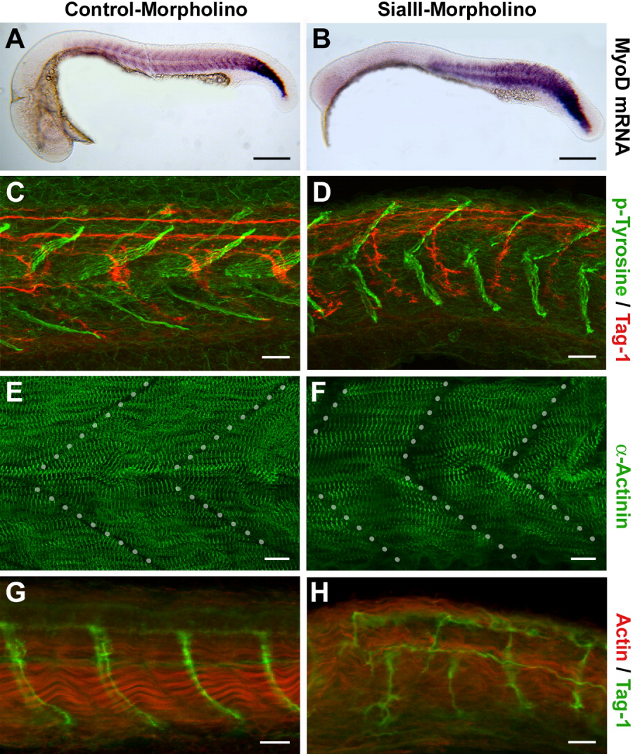
Fig. 4 Knockdown of St8SiaIII induces malformations of myotomes and an abnormal growth pattern of motoneurons in developing zebrafish. A,C,E,G: Embryos injected with control morpholinos: (A, C, E) GeneTools control morpholino, (G) PST-morpholino (Marx et al., 2007). B,D,F,H: Embryos injected with St8SiaIII-specific morpholino (2 ng/μl). All embryos are shown rostral to the left. A, B: In situ hybridization using a mytome-specific probe (MyoD) shows a reduction of brain structures in morpholino-injected embryos at 25 hpf. C-F: Embryos at 33 hpf after morpholino knockdown immunolabelled for (C, D) phospho-Tyrosine (green) and the axonal marker Tag-1 (red) or (E, F) α-Actinin (green). Dotted lines in E and F indicate the transversal mysepta. Morphants are characterized by shortened myotomes and myosepta forming a more obtuse angle than in the controls. G,H: Embryos at 35 hpf after morpholino knockdown were immunolabelled for Actin (red) and the axonal marker Tag-1 (green). Muscle fibrils lose their well-ordered bundling, and motoraxons grow in a disordered pattern. Scale bars = 200 μm (A, B), 20 μm (C, D, G, H), 10 μm (E, F).