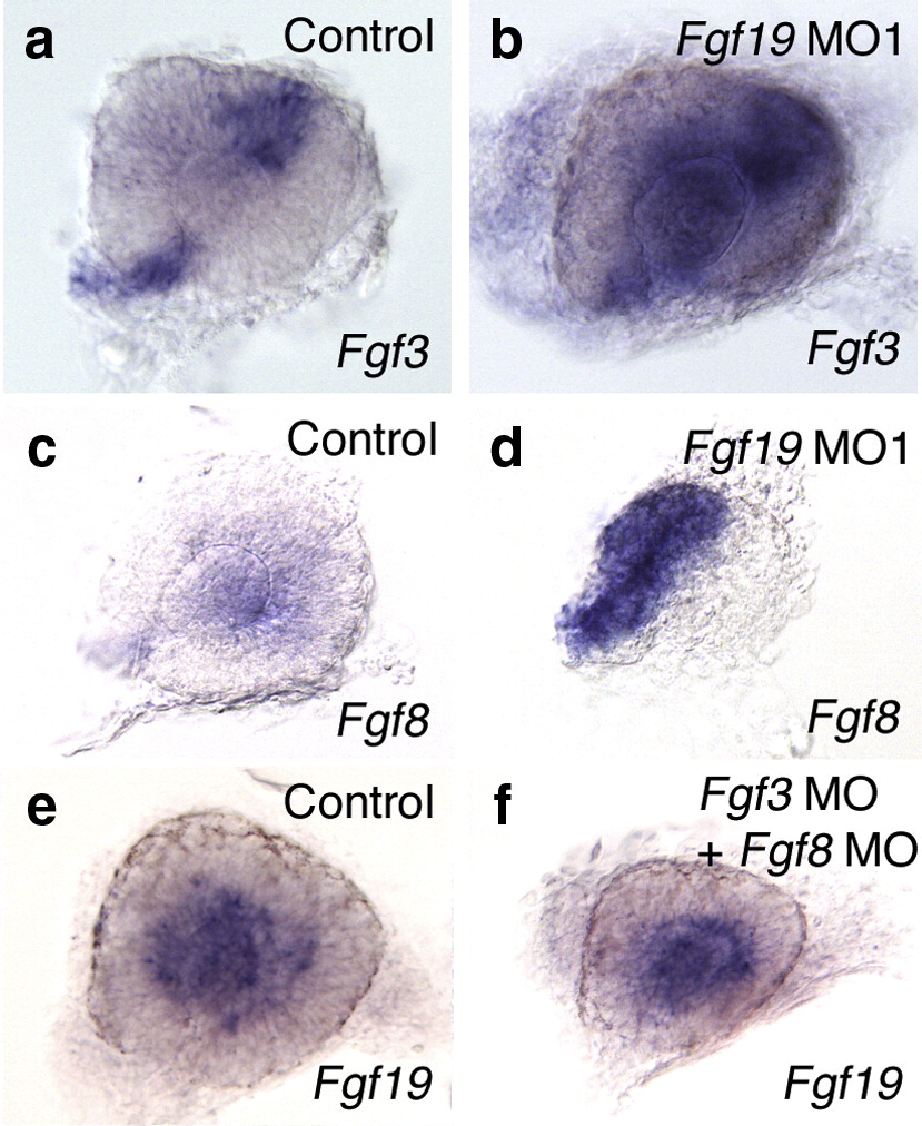
Fig. 9 Interactions between Fgf3, Fgf8 and Fgf19 in the eye. (a, b) The expression of Fgf3 in control and Fgf19 MO1-injected embryos at 24 hpf. In control embryos, the expression of Fgf3 was detected in the optic stalk and the dorsal retina. The expression of Fgf3 was up-regulated in the retina of Fgf19 MO1-injected embryos. (c, d) The expression of Fgf8 in control and Fgf19 MO1-injected embryos at 24 hpf. In control embryos, the expression of Fgf8 was detected in the proximal retina. The ectopic expression of Fgf8 was detected in the nasal retina of Fgf19 MO1-injected embryos. (e, f) The expression of Fgf19 in wild-type embryos and embryos injected with both Fgf3 MO and Fgf8 MO at 24 hpf. In embryos injected with both Fgf3 MO and Fgf8 MO, Fgf19 expression in the inner retina was slightly decreased, while that in the lens was unaffected. Lateral views with anterior to the left and dorsal to the top.
Reprinted from Developmental Biology, 313(2), Nakayama, Y., Miyake, A., Nakagawa, Y., Mido, T., Yoshikawa, M., Konishi, M., and Itoh, N., Fgf19 is required for zebrafish lens and retina development, 752-766, Copyright (2008) with permission from Elsevier. Full text @ Dev. Biol.

