
|
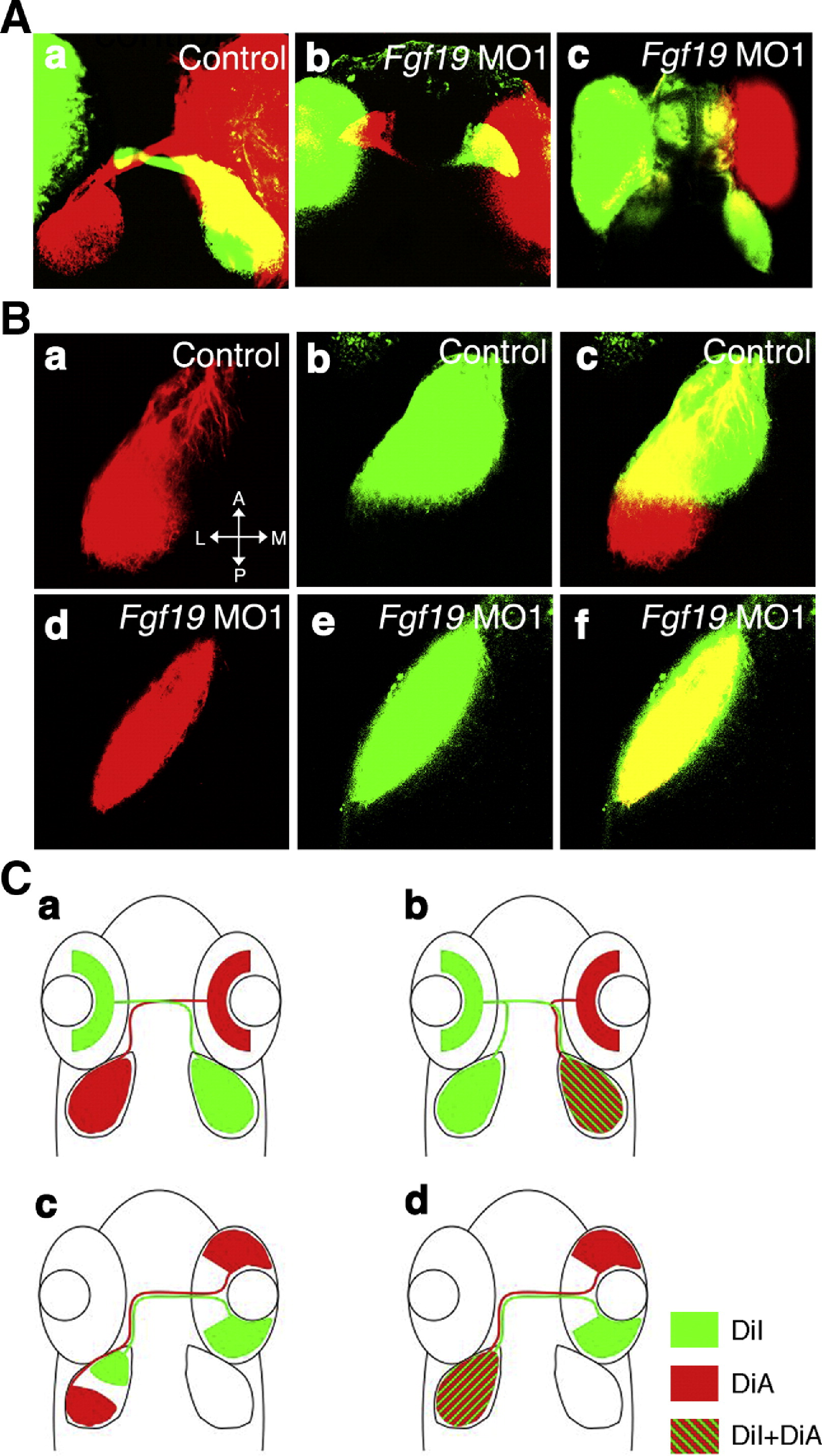
Fig. 8 Guidance of retinal ganglion cell axons in Fgf19 MO1-injected embryos. A: Embryos were injected with control MO (a) and Fgf19 MO1 (b, c). (a) In control embryos, retinal axons projected to contralateral optic tecta. (b) The mild phenotype embryos showed normal projections. (c) In the severe phenotype embryos, retinal axons frequently projected to ipsilateral optic tecta or in some cases projected to both ipsilateral and contralateral optic tecta. B: Embryos were injected with control MO (a?c) and Fgf19 MO1 (d?f). Retinal ganglion cell axons anterogradely double-labeled by DiI (green) and DiA (red) in the dorsotemporal and dorsonasal quadrants. (a?c) In control embryos, nasal and temporal retinal ganglion cells projected to the posterior and anterior tectum, respectively. (d?f) Both nasal and temporal retinal ganglion cells projected to the entire tectum in Fgf19 MO1-injected embryos. C: Schematics of guidance of retinal ganglion cell axons in control (a, c) and Fgf19 MO1-injected (b, d) embryos. Dorsal views with anterior to the top.
Reprinted from Developmental Biology, 313(2), Nakayama, Y., Miyake, A., Nakagawa, Y., Mido, T., Yoshikawa, M., Konishi, M., and Itoh, N., Fgf19 is required for zebrafish lens and retina development, 752-766, Copyright (2008) with permission from Elsevier. Full text @ Dev. Biol.