
IMAGE
Fig. 2
Image
|
Figure Caption
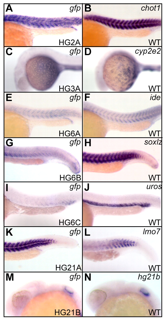
Fig. 2 Expression patterns of genes at the integration sites. (A,C,E,G,I,K,M) Whole-mount in situ hybridization of embryos heterozygous for respective insertions (bottom right) at 24 hpf using the gfp probe. (B,D,F,H,J,L,N) Whole-mount in situ hybridization of wild-type embryos at 24 hpf using probes indicated (top right). Signals were detected in myotome (A,B), yolk (C,D), myotome (E-H), ventral mesoderm (I,J), myotome (K,L) and otic vesicle (M,N).
Figure Data
Acknowledgments
This image is the copyrighted work of the attributed author or publisher, and
ZFIN has permission only to display this image to its users.
Additional permissions should be obtained from the applicable author or publisher of the image.
Full text @ Development