
|
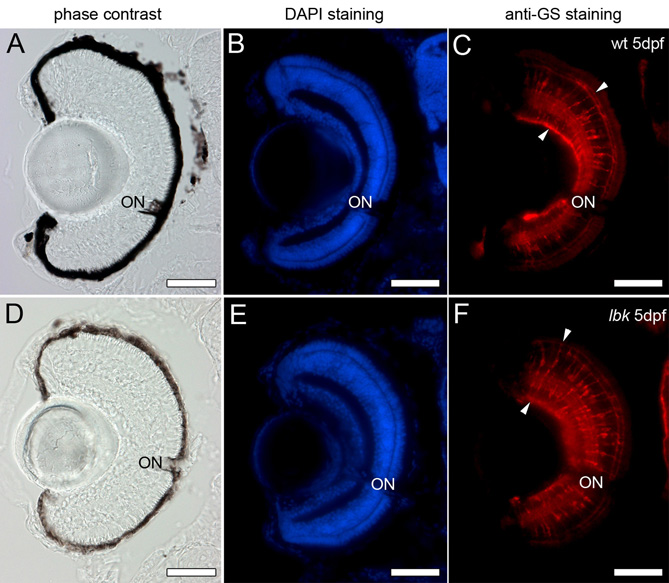
Fig. S6 Retinal Müller glia cells show a normal morphology in lbk mutants. Cryosections of 5 dpf wild-type and lbk eyes were stained with DAPI (blue; panels B and E) to show the position of the nuclei in the retina and an antibody against the glia-specific enzyme glutamine synthetase to label the retinal Müller glia cells (red; arrowheads in C and F). Phase-contrast images of the sections of wild-type and lbk eyes are shown in A and D, respectively. No obvious differences in the number, arrangement and morphology of the Müller glia cells were observed between wild-type and lbk larvae. ON, optic nerve. Scale bars: 50 μm.