Image
Figure Caption
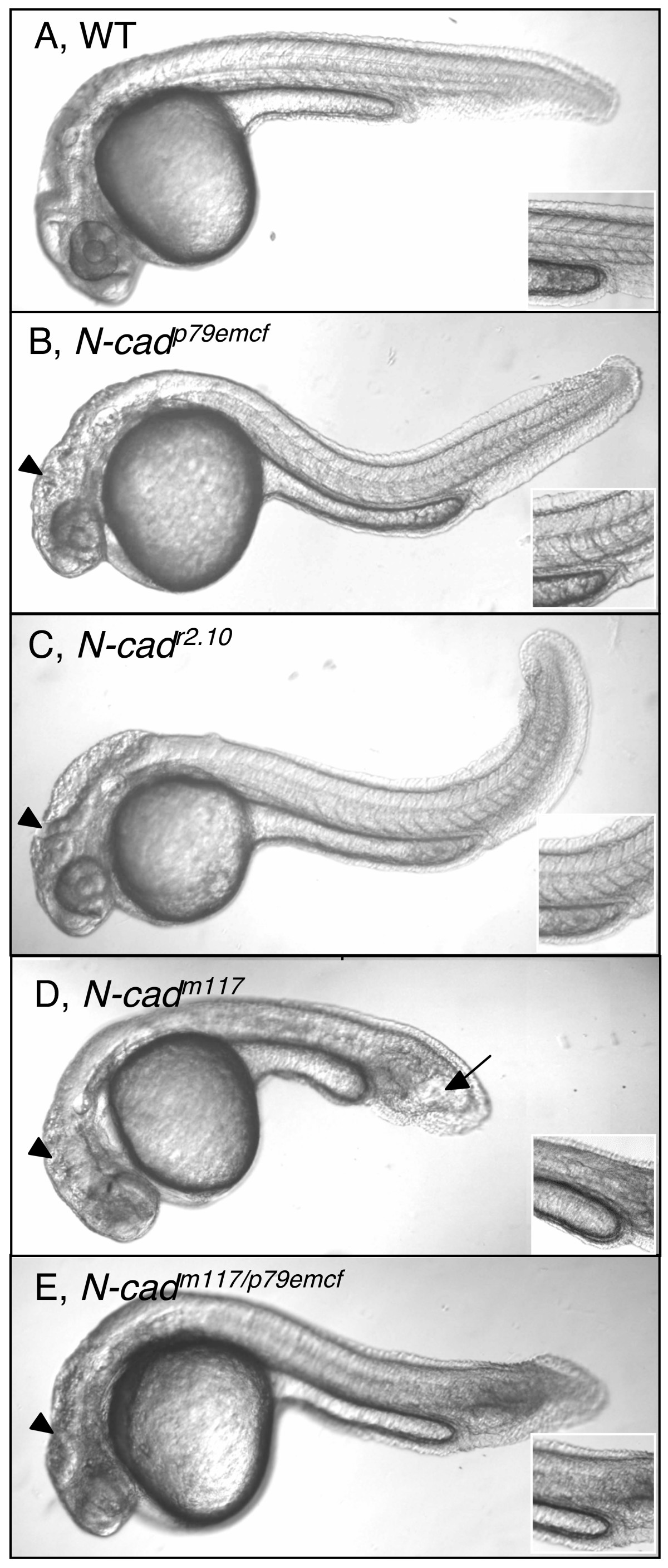
Fig. 1 Loss of N-cadherin function causes axis shortening. (A-E) Lateral views of live 30 hpf zebrafish embryos imaged with Nomarski optics. Anterior is to the left, dorsal is up. Insets show images of somites at the level of the yolk sac extension. Black arrowheads point to brain defects observed in N-cad mutants. Black arrow points to the characteristic club-shaped, shortened tail in N-cadm117 homozygous mutants. WT (A), N-cadp79emcf homozygous mutant (B), N-cadr2.10 homozygous mutant (C), N-cadm117 homozygous mutant (D), N-cadm117/p79emcf transheterozygote mutant (E) embryos.
Figure Data
Acknowledgments
This image is the copyrighted work of the attributed author or publisher, and
ZFIN has permission only to display this image to its users.
Additional permissions should be obtained from the applicable author or publisher of the image.
Full text @ BMC Dev. Biol.

