
|
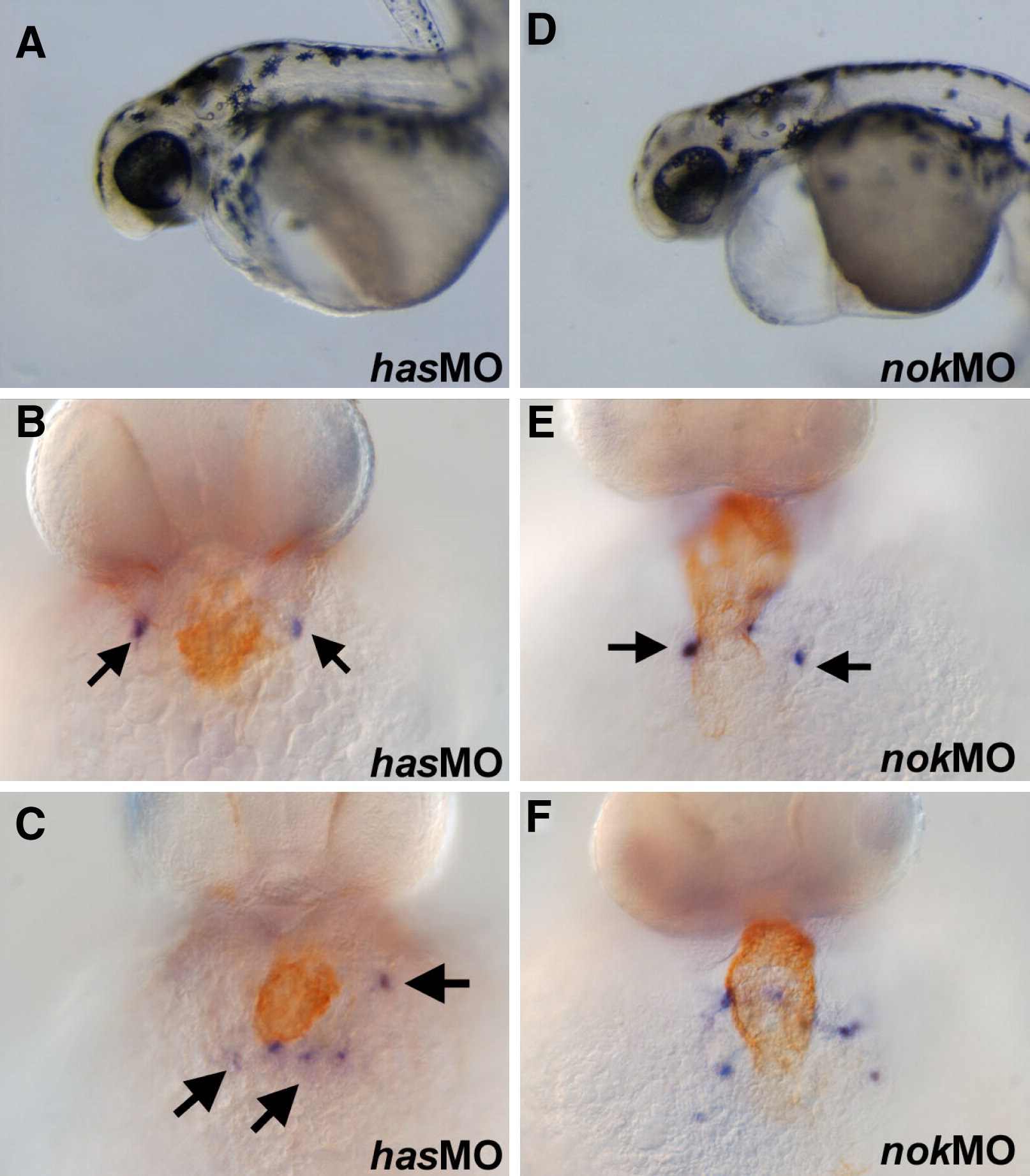
Fig. 7 The cell polarity genes has and nok are required for proper PEO formation. (A) hasMO embryos shown live at 48 hpf. (B) hasMO embryo stained with MF20 (brown) and wt1 (blue) displays a midline heart but bilateral clusters of wt1 cells (arrows). (C) An example of a hasMO embryo displaying scattered wt1-positive cells (arrows) adjacent to the heart. (D) 48 hpf embryos injected with nokMO displaying characteristic pericardial edema. (E and F) nokMO embryo at 48 hpf stained with wt1 (blue) and MF20 (brown). Note the midline heart in but the bilateral spots of wt1 expression (arrows in panel E) and scattered wt1-positive cells in panel F.
Reprinted from Developmental Biology, 315(1), Serluca, F.C., Development of the proepicardial organ in the zebrafish, 18-27, Copyright (2008) with permission from Elsevier. Full text @ Dev. Biol.