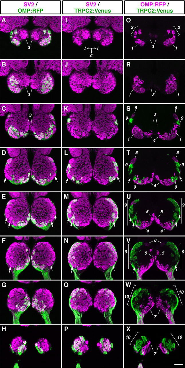
Fig. 6 Mutually exclusive glomerular innervation by two types of OSNs. Horizontal sections through the OB of OMP:RFP; TRPC2:Venus adult zebrafish were stained with anti-RFP, anti-GFP, and anti-SV2 antibodies. A-H, Merged images of staining for RFP (green) and SV2 (magenta). I-P, Merged images of staining for Venus (green) and SV2 (magenta). Q-X, Merged images of staining for RFP (magenta) and Venus (green). The alternate eight sections from a series of OB sections are ordered from the dorsalmost (top) to the ventralmost (bottom) section. All photographs represent stacked images of optical sections. Arrows indicate glomeruli that were not labeled with either RFP or Venus. Numbers for single glomeruli or glomerular subgroup: 1, dorsal cluster; 2, dorsal cluster-associated glomeruli; 3, mediodorsal posterior glomeruli; 4, anterior plexus; 5, medial glomeruli; 6, medioventral posterior glomerulus; 7, ventromedial glomeruli; 8, lateroposterior glomerulus; 9, lateral chain; 10, ventrolateral glomeruli. Posterior is at the top of each panel. a, Anterior; l, lateral. Scale bar, 100 μm.
Image
Figure Caption
Acknowledgments
This image is the copyrighted work of the attributed author or publisher, and
ZFIN has permission only to display this image to its users.
Additional permissions should be obtained from the applicable author or publisher of the image.
Full text @ J. Neurosci.

