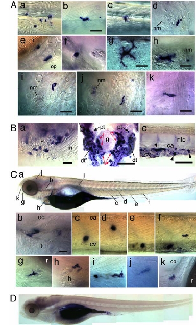
Fig. S5 Caged-Fluorescein-Dextran-Based Cell-Tracing Data Supplemental to Figures 4 and 5. (A) Single labelled cells outside hematopoietic tissues in the embryos shown in Figure 4A (af), 4E (g,h) and 5C (i-k); (a-c) in the trunk, beneath the epidermis at or near the horizontal septum; (d,h) At the base of a cephalic (d) or caudal (h) neuromast (nm); (e) Olfactory pit (op); (f) Gills; (g) Anal fin epidermis; (i-k) Ventral head; (B) Labelled cells in the right thymus, ventral view rostral left (a), pronephros, red arrows, dorsal view rostral up (b), and tail, lateral view rostral left (c) of an embryo uncaged in the trunk DA/PCV joint at 3 dpf, immunostained at 5 dpf. (C) Labelled cells found at 5 dpf following uncaging of ten primitive macrophages in the yolk sac at 1 dpf. (b) the labelled macrophage closest to the unlabelled thymus is in contact with the otic epithelium. (c-f) Labelled large (stromal) macrophages in the CHT; (g) mesenchyme rostral to the eye; (h) heart; (i) horizontal septum; (j) brain parenchyme (primitive microglia); (k) olfactory pit. (D) Control embryo, injected with caged-fluorescein dextran and immunostained for fluorescein at 5 dpf with no prior UV-induced uncaging. Only the gut is labelled. R, retina; t, thymus; m, muscle; g, glomerulus; dt, distal tubules; pt proximal tubules. Bars: (A), (B-a), (C), 20 μm; (B-b,c), 50 μm.
Reprinted from Immunity, 25(6), Murayama, E., Kissa, K., Zapata, A., Mordelet, E., Briolat, V., Lin, H.F., Handin, R.I., and Herbomel, P., Tracing Hematopoietic Precursor Migration to Successive Hematopoietic Organs during Zebrafish Development, 963-975, Copyright (2006) with permission from Elsevier. Full text @ Immunity

