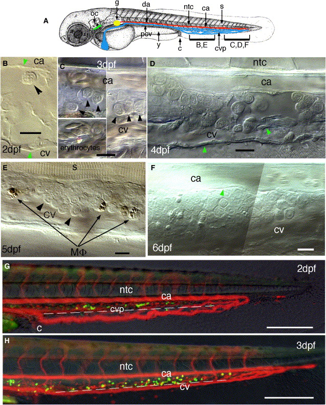
Fig. 1 In Vivo Observation of Hematopoietic Progenitors in the Ventral Tail of Zebrafish through the First Week of Development (A) Anatomical landmarks used in this study: dorsal aorta (da), posterior cardinal vein (pcv), cloaca (c), caudal artery (ca), caudal vein plexus (cvp), thymus (green), pronephric glomerulus (g, yellow), notochord (ntc), somite muscles (s), otic capsule (oc). (B?F) Video-enhanced DIC microscopic observation in the ventral tail, showing progressive accumulation of hematopoietic progenitors (a few shown by black arrowheads) between the CA and definitive CV; (B) 2 dpf; (C) 3 dpf; the lower left panel shows for comparison, both in flat and profile view, the distinct shape of primitive erythrocytes past 2.5 dpf, with a very small nucleus (3.5 μm) and flat elliptical cytoplasm; (D) 4 dpf; (E) 5 dpf; (F) 6 dpf. Green arrowheads, vascular endothelium; Mφ, macrophages. (G and H) DIC, red and green fluorescence overlay images of a gata1-dsred, CD41-gfp double transgenic embryo at (G) 2 dpf and (H) 3 dpf; a dotted line indicates the ventral limit of somite muscles; circulating DsRed+ primitive erythrocytes delineate the blood flow. All lateral views, rostral to the left. Scale bars represent 10 μm in (B)?(F), 100 μm in (G) and (H).
Reprinted from Immunity, 25(6), Murayama, E., Kissa, K., Zapata, A., Mordelet, E., Briolat, V., Lin, H.F., Handin, R.I., and Herbomel, P., Tracing Hematopoietic Precursor Migration to Successive Hematopoietic Organs during Zebrafish Development, 963-975, Copyright (2006) with permission from Elsevier. Full text @ Immunity

