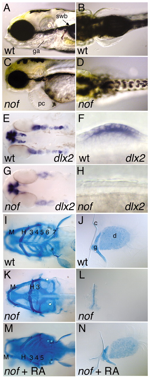
Fig. 3 Phenotype of nof homozygotes; wt: wild-type sibling. In A-I,K,M, anterior is to the left; in J,L,N proximal is to the left. (A,C) Lateral, (B,D) dorsal, (E-N) ventral views. (A-D) Larvae on day 5; ga, gill arches; pc, pericardial cavity; swb, swim bladder. (E,G) Branchial arch primordium of 36 hours embryos. (F) Presence and (H) absence of pectoral fin bud and AER marker dlx2 at 28 hours. (I,K) Cartilage pattern in heads and (J,L) fins of day 5 larvae. M, mandibular and H, hyoid arches; 3-7, gill arches; c, cleithrum; d, distal fin skeleton; g, proximal pectoral girdle. (M) Rescue of arch cartilages and (N) pectoral fins by treatment of nof homozygotes with 10–9 M retinoic acid at 30% epiboly until 16 hours.
Image
Figure Caption
Figure Data
Acknowledgments
This image is the copyrighted work of the attributed author or publisher, and
ZFIN has permission only to display this image to its users.
Additional permissions should be obtained from the applicable author or publisher of the image.
Full text @ Development

