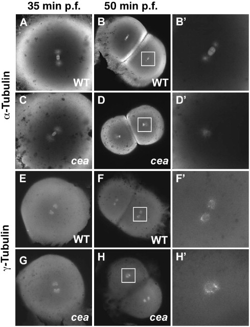
Fig. 2 Spindle organization and centrosome duplication defects in maternally mutant cea embryos. (A–D′) Fixed wild-type (A, B′) and mutant (C, D′) embryos labeled with anti-α-tubulin antibody. Spindle organization is normal in the mutant immediately prior to the first cell division (35 min p.f.; panel C; compare to panel A), but is defective in a fraction of blastomeres in the next cleavage cycle (50 min p.f.; panels D, D′; compare to panels B, B′). (E–H′) Fixed wild-type (E, F′) and mutant (G, H′) embryos labeled with anti-γ-tubulin antibody. Centrosome duplication appears normal immediately prior to the first cleavage division (35 min p.f.; (G), compare to (E)) but is defective in a fraction of the blastomeres in the following cleavage cycle (50 min p.f.; panels H, H2, compare to panels F, F′). Animal views. Panels B′, D′, F′, H′ are enlargements of the area indicated by the squares in panels B, D, F, H, respectively.
Reprinted from Developmental Biology, 312(1), Yabe, T., Ge, X., and Pelegri, F., The zebrafish maternal-effect gene cellular atoll encodes the centriolar component sas-6 and defects in its paternal function promote whole genome duplication, 44-60, Copyright (2007) with permission from Elsevier. Full text @ Dev. Biol.

