
|
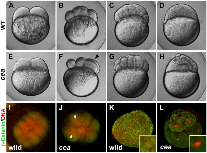
Fig. 1 Phenotype of maternally mutant cea embryos. (A?H) Side views of live wild-type (A?D) and mutant (E?H) embryos at the following time points: (A, E) 45 min post fertilization (p.f.), (B, F) 1.25 h p.f., (C, G) 2.25 h p.f., (D, H) 3 h p.f. (corresponding, in wild-type, to the 2-, 8-, 128- and 1000-cell stages, respectively). Maternally mutant cea embryos show a normal first cell division (E) but fail in a fraction of subsequent divisions (F, arrowhead), leading to blastula with irregularly sized blastomeres (G) and largely syncytial embryos (H). (I?L) Animal views of fixed wild-type (I, K) and mutant (J, L) embryos labeled to detect β-catenin (green) and DNA (red). At 1.25 h p.f. (8-cell stage in wild-type; panels I, J), the mutant embryo reveals a failure of DNA and cell division in a fraction of the blastomeres (arrowheads indicate nuclei of cells that did not undergo division). At 3 h p.f. (1000-cell stage in wild-type; panels K, L), mutant embryos exhibit enlarged nuclei (inset in panel L, compare to panel K).
Reprinted from Developmental Biology, 312(1), Yabe, T., Ge, X., and Pelegri, F., The zebrafish maternal-effect gene cellular atoll encodes the centriolar component sas-6 and defects in its paternal function promote whole genome duplication, 44-60, Copyright (2007) with permission from Elsevier. Full text @ Dev. Biol.