
|
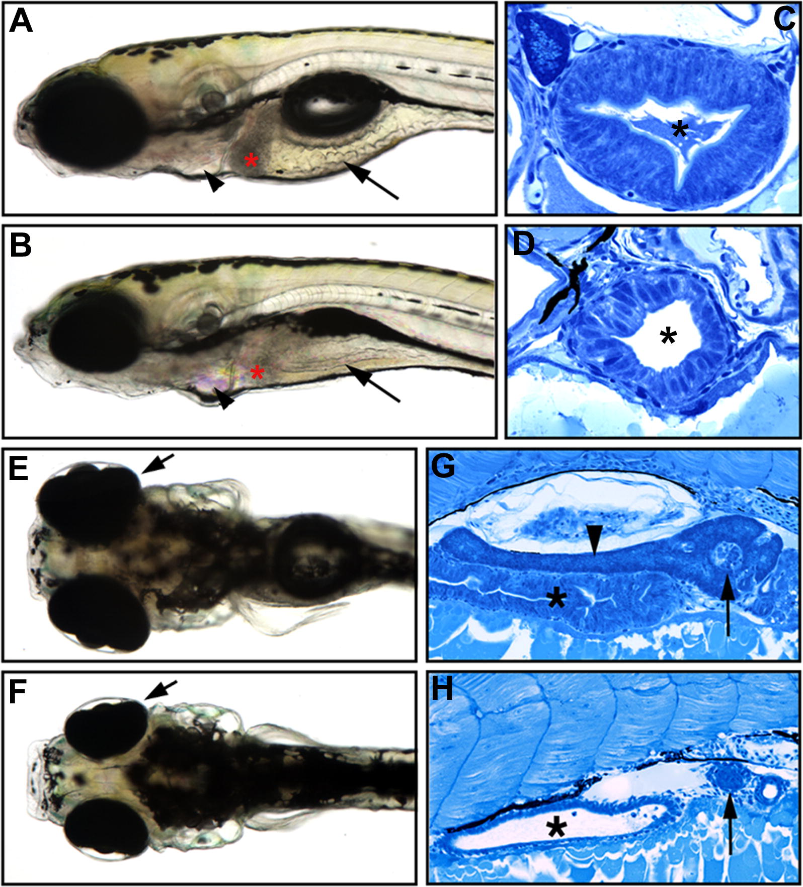
Fig. 1 The slj Mutation Disrupts Intestinal and Exocrine Pancreas Development (A?D) Lateral views of 5-dpf wild-type (A) and slj (B) larvae, with representative histological cross-sections (C and D). The slj intestine is small, thin walled, and lacks folds (arrows, [A and B]) compared with wild type, and the slj terminal branchial arches are reduced (arrowheads). The columnar morphology and apical microvilli of the slj epithelium are less developed than in wild-type larvae (C and D); an asterisk (*) indicates the intestinal lumen. (E and F) The size of the slj liver (indicated by red asterisk in [A] and [B]) and the retinae (arrows, [E and F], dorsal view) are also reduced. (G and H) Although exocrine tissue is well developed in wild-type embryos (arrowhead in [G]), it is markedly reduced or absent in slj embryos (H); asterisks indicate intestine. By contrast, the slj and wild-type pancreatic islets are of comparable size (arrows).