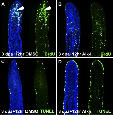
Fig. S4 ActβA Signaling Is Required for Blastemal Proliferation. (A?D) Longitudinal sections of fin rays at 3.5 dpa. In control fins, the blastema (arrowheads) displays a massive proliferation as visualized by BrdU incorporation in green (A) and only sparse mesenchymal cells labeled by TUNEL cell-death assay in green (C). Fins regenerating in normal conditions for 3 days and then transferred to water with Alk-i for the next 12 hr display a reduction of blastemal proliferation (B). No enhancement in mesenchymal cell death is present after the drug shift (D). Cell death is detected in the outer layers of the epidermis. (E and F) Quantification of BrdU-positive cells in the epidermis and in the mesenchyme after inhibitor treatment for 24 hpa starting immediately after amputation (E) and after the drug shift (3 days at normal conditions, followed by the next 6, 12 or 24 hr with Alk4-i (F). The values are presented as a percentage of the control samples, which were set as 100%. Error bars represent the SEM. n = 10 (two or three representative sections from four fins were analyzed). *p < 0.01 indicates a significant difference between inhibitor-treated fins and the control.
Image
Figure Caption
Acknowledgments
This image is the copyrighted work of the attributed author or publisher, and
ZFIN has permission only to display this image to its users.
Additional permissions should be obtained from the applicable author or publisher of the image.
Full text @ Curr. Biol.

