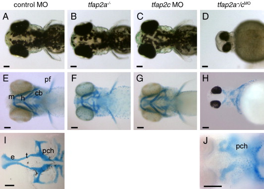
Fig. 3 tfap2a/c deficient embryos display non-additive defects in pigmentation and jaw morphology. (A?D) Dorsal views of live 72 hpf embryos. (A) Melanophores are abundant on the dorsal aspect of the head in a control-MO injected wild-type embryo, slightly reduced in panel B, a control MO-injected tfap2a mutant, normal in panel C, a tfap2c MO-injected wild-type embryo, but absent from panel D, a tfap2a-/cMO embryo (6 of 6 tfap2a-/cMO embryos). (E?H) Ventral views of 4 dpf embryos processed to reveal cartilage (alcian green), and bleached to remove pigment (except panel H). (E) Cartilages derived from mandibular (1st arch, m), hyoid (2nd arch, h), and ceratobranchial (3rd?7th arches, cb) arches are seen in a control embryo. Pectoral fin (pf) is also visible. (F) A control MO-injected tfap2a homozygous mutant embryo, with normal sized mandibular, but severely reduced hyoid and ceratobranchial cartilages, as previously reported (Schilling et al., 1996). (G) A tfap2c MO-injected wild-type embryo, in which all craniofacial cartilages appear normal. (H) A tfap2a-/cMO embryo, in which ventral craniofacial cartilage elements and pectoral fin are absent (10 of 10 tfap2a-/cMO embryos). (I) Ventral view of dissected neurocranium from a control embryo. (J) Ventral view of a tfap2a-/cMO embryo, shown at higher magnification, only the mesoderm-derived posterior neurocranium is visible (10 of 10 tfap2a-/cMO embryos). e, ethmoid plate; pch, parachordal; t, trabeculae cranii (Schilling et al., 1996). Scale bars: 100 μm.
Reprinted from Developmental Biology, 304(1), Li, W., and Cornell, R.A., Redundant activities of Tfap2a and Tfap2c are required for neural crest induction and development of other non-neural ectoderm derivatives in zebrafish embryos, 338-354, Copyright (2007) with permission from Elsevier. Full text @ Dev. Biol.

