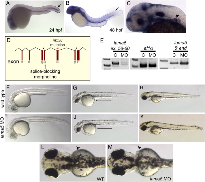
Fig. 3 lama5 MO-injected embryos recapitulate the lama5 mutant phenotype. (A?C) By in situ hybridization, lama5 is expressed in the developing fin fold epidermis (arrows) at 24 hpf (A) and 48 hpf (B) and the developing pectoral fin (arrowhead) at 36 hpf (C). (D) The lama5 splice-blocking MO targets the exon 58 splice donor (blue arrow), one exon upstream of the m538 mutation (orange asterisk). (E) Injection of the lama5 MO successfully blocks splicing of the lama5 transcript (C, uninjected; MO, 3 ng lama5 MO; PCR primers are shown in green in panel D), but does not affect the control ef1α transcript or the 52 end of the lama5 transcript, demonstrating that the lama5 transcript is not eliminated by non-sense-mediated decay in MO-injected embryos. (F?K) Lama5 MO-injected embryos (I?K) display fin fold defects and yolk extension abnormalities (J, bracket) compared to uninjected controls (F?H) at 28 hpf (F, I), 48 hpf (G, J) and 72 hpf (H, K). (L, M) Pectoral fins at 72 hpf (arrowheads). In the morphants, the pectoral fins are abnormal (M) compared to those in wild-type embryos where the pectoral fins lie flat against the yolk (L). All images are lateral views with anterior to the left, except panels L and M, which show dorsal views with anterior to the left.
Reprinted from Developmental Biology, 311(2), Webb, A.E., Sanderford, J., Frank, D., Talbot, W.S., Driever, W., and Kimelman, D., Laminin alpha5 is essential for the formation of the zebrafish fins, 369-382, Copyright (2007) with permission from Elsevier. Full text @ Dev. Biol.

