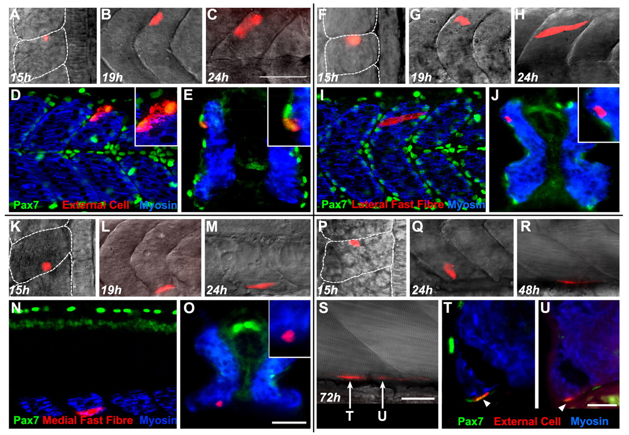
Fig. 2 Distinct fates of ABCs and posterior cells. (A-E) ABCs give rise to external cells. (A) The injected ABC (red). (B) Four hours later, this ABC had given rise to a cell on the external somite surface. (C) Ten hours after injection, 2 cells remain on the surface of the somite. (D) Both cells (red) are Pax7-positive [colocalisation of rhodamine and Pax7 (green) in the nucleus appears yellow] on the surface of the myotome (blue). (E) Transverse section of one labeled cell shown in D. (F-J) ABCs give rise to muscle. (F) The injected ABC (red). (G) Four hours later, this cell is on the external somite surface. (H) Ten hours after injection, this ABC formed an elongated fibre. (I) The elongated muscle fibre (red) is obliquely oriented, like fast fibres, Pax7-negative (green), and is superficial within the myotome (blue). (J) Transverse section of the embryo in I, the labeled cell is a lateral fast fibre. (K-O) Posterior cells elongate early. (K) The injected posterior cell. (L) Four hours later, the posterior cell had elongated, deep within the somite. (M) Ten hours after injection, the posterior cell had formed an elongated muscle fibre. (N) The elongated fibre (red) is Pax7-negative (green), and is deep within the myotome (blue). (O) Transverse section of the embryo in N, the labeled cell is a very medial fast fibre. (P-U) External cells persist into the larval stage. (P) The injected ABC (red). (Q) Ten hours after injection, this cell is on the external surface of the myotome. (R) Thirty-four hours after injection, this cell had generated multiple cells. (S) Fifty-seven hours after injection, all injected cells remain on the external surface, arrows show the position of sections shown in T,U. (T,U) The injected cell gave rise to two Pax7-positive (yellow) cells on the surface of the myotome (blue). A,F,K and P are dorsal views, anterior to the top, the notochord is at the right of the image. B-D,G-I,L-N and Q-S are lateral views, with anterior to the left and dorsal to the top. E,J,O,T,U are transverse sections with dorsal to the top. Scale bars: 50 μm in C for A-C,F-H,K-M,P-R; 50 μm in O for D,E,I,J,N,O; 25 μm in S; 25 μm in U for T,U.
Image
Figure Caption
Figure Data
Acknowledgments
This image is the copyrighted work of the attributed author or publisher, and
ZFIN has permission only to display this image to its users.
Additional permissions should be obtained from the applicable author or publisher of the image.
Full text @ Development

