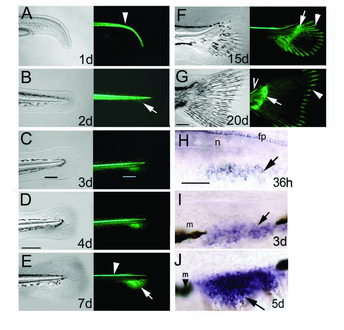
Fig. 1 The ontogeny of the caudal and pectoral fins is marked by continuous GFP expression in the transgenic zebrafish line 2.2shh:gfp:ABC#15. A-G, The first 20 days of zebrafish caudal fin development. Bright field view of caudal fin on left panels, right panels show fluorescence signals of GFP activity. A, GFP in the notochord is shown by an arrowhead. B, GFP protein is detected from 2 days onwards in the embryonic fin fold mesenchyme. C, D, GFP expression indicates that the caudal fin mesenchyme occupies the gap of the melanophore streak at 3?4 dpf (black and white bars in C). E, GFP in the caudal fin primordium is split anteroposteriorly at 7 dpf (arrow at boundary of domains). Notochord expression is diminished, while floor plate expression remains active (arrowhead). F, G: GFP expression extends caudally and tilts dorsally indicating the formation of adult caudal fin morphology with dorsal and ventral lobes. In late larval development proximal GFP expression is present in the endoskeletal territory (arrows in F, G) and distally in the fin rays (arrowheads in F, G). GFP is expressed continually in the floor plate (open arrowhead in G). H-J, gfp mRNA expression in the ACFP is first detected at 1.5 dpf by in situ hybridization (H) then extends ventrally (arrows in I, J) between the melanophores (arrowhead with m). Age of larvae developing at 28°C is indicated in days post fertilization (d). Scale bar in D represents 200 μm (for panels A-F) and 100 μm in G. Scale bar in H represents 100 μm (H-J). Abbreviations; fp, floor plate, n, notochord, m, melanophore.
Image
Figure Caption
Figure Data
Acknowledgments
This image is the copyrighted work of the attributed author or publisher, and
ZFIN has permission only to display this image to its users.
Additional permissions should be obtained from the applicable author or publisher of the image.
Full text @ BMC Dev. Biol.

