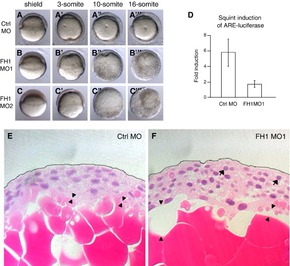
Fig. 1 FoxH1 depletion phenotype. (A?C) Two independent FoxH1 MOs produce similar phenotypes. 8 ng of the indicated MO was injected into wild type embryos whose chorions were removed before being mounted in soft agar/agarose and simultaneously time lapse documented, using a lateral view. Dorsal is to the right in panel A, and unknown in panels B, C. Primes (2) indicate different time points for the same embryos. (D) FoxH1 MO1-injected embryos show reduced FoxH1 activity in response to Squint. Wild type embryos were injected with 10 pg of squint mRNA and 25 pg of reporter plasmids, plus 8 ng of either control (Ctrl) MO or FoxH1 (FH1) MO1. Injected embryos were incubated to the dome stage and then lysed for the luciferase assay. (E?F) Histology of a FoxH1 morphant.Wild type embryos were injected with 8 ng of either control or FoxH1 morpholino, fixed at 8 hpf when control embryos were at the 70% epiboly stage and paraffin mounted prior to sectioning and hematoxylin and eosin staining. Arrowheads point to gaps between the yolk and embryonic cells that are plentiful in morphants. (F). Arrows point to rounded nuclei that are abundant in morphant (F), but not control (E) embryos. Abbreviations: FH1, FoxH1; ctrl, control.
Reprinted from Developmental Biology, 310(1), Pei, W., Noushmehr, H., Costa, J., Ouspenskaia, M.V., Elkahloun, A.G., and Feldman, B., An early requirement for maternal FoxH1 during zebrafish gastrulation, 10-22, Copyright (2007) with permission from Elsevier. Full text @ Dev. Biol.

