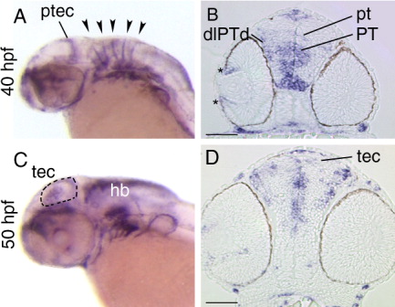
Fig. 7 Expression of sema3Gb. A,C: Lateral views of whole-mount embryos. B,D: Stage-matched transverse sections. A: At 40 hpf, in situ signal is evident in the posterior tectum and the rhombomeric boundaries (arrowheads). B: At 40 hpf, sema3Gb is expressed in the peripheral retina (asterisk). Several areas of the diencephalon express sema3Gb: dorsolateral region of the dorsal posterior tuberculum, the dorsoventral axis of the posterior tuberculum, and the pretectal area. C,D: At 50 hpf, a subset of cells dispersed throughout the tectum express sema3Gb, which is evident in the whole-mount embryo (dashed line outlines the tectum) and in the section. pt, pretectum; ptec, posterior tectum; tec, tectum; PT, posterior tuberculum; dlPTd, dorsolateral region of the dorsal posterior tuberculum; Scale bars in B,D = 50 μm.
Image
Figure Caption
Figure Data
Acknowledgments
This image is the copyrighted work of the attributed author or publisher, and
ZFIN has permission only to display this image to its users.
Additional permissions should be obtained from the applicable author or publisher of the image.
Full text @ Dev. Dyn.

