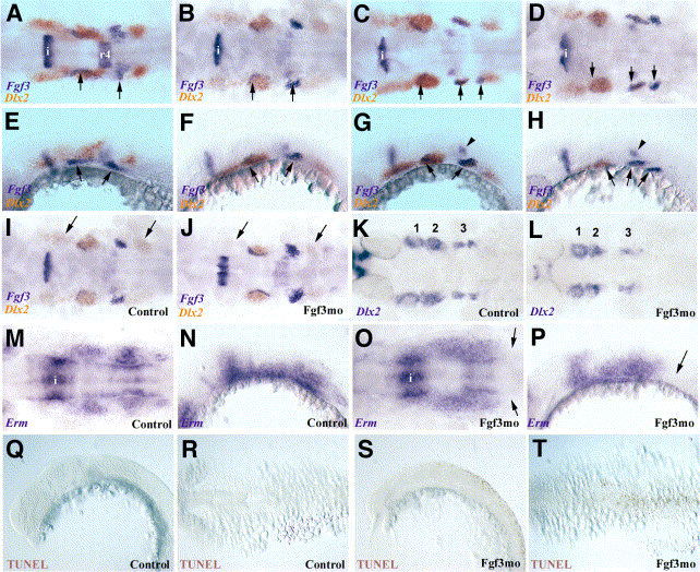
Fig. 4 Fgf3 expression by pharyngeal pouch endoderm closely associated with neural crest streams. (A?H) Close relationship of endodermal Fgf3 expression to neural crest streams shown by hybridisation for Fgf3 (blue) and Dlx2 (orange) transcripts. Dorsal (A?D) and lateral (E?H) views of 18 (A, E), 21 (B, F), 24 (C, G), and 28 hpf (D, H) embryos. Fgf3 is expressed in endoderm of the first, second, and third pharyngeal pouches (arrows), also the isthmus (i) and rhombomere 4 (r4) in younger embryos (A, B, E, F) and in the anterior of the otic vesicle in older embryos (B?D, F?H; arrowhead in G, H). (I?L) Dlx2-positive neural crest cells are still present in embryos at 21 and 30 hpf following inhibition of Fgf3 translation with morpholinos. In situ hybridisation for Dlx2 (orange) and Fgf3 (blue) transcripts in embryos at 21 hpf injected with a control morpholino (I; dorsal view) or Fgf3mo (J; dorsal view). Arrows indicate first and third neural crest streams. Dlx2 expression at 30 hpf in embryos injected with a control morpholino (K; dorsal view) or Fgf3mo (L; dorsal view). (M?P) Posterior but not anterior expression of Erm by neural crest cells in 18-hpf embryos is reduced following inhibition of Fgf3. Dorsal (M, O) and lateral (N, P) views of embryos injected with control morpholino (M, N) or Fgf3mo (O, P). Erm transcripts are not detected in the most posterior of the neural crest streams (arrows). Isthmic Erm expression (i) is indicated in (M) and (O). (Q?T) Cell death is not detected in migrating crest streams at 20 hpf following inhibition of Fgf3. Dorsal (Q, S) and lateral (R, T) views of embryos injected with control (Q, R) or Fgf3mo oligonucleotides and analysed by TUNEL staining. No apoptotic cells are apparent in control embryos (Q, R), while death is restricted to dorsal neural tube at hindbrain and cord levels following inhibition of Fgf3 (S, T).
Reprinted from Developmental Biology, 264(2), Walshe, J. and Mason, I., Fgf signalling is required for formation of cartilage in the head, 522-536, Copyright (2003) with permission from Elsevier. Full text @ Dev. Biol.

