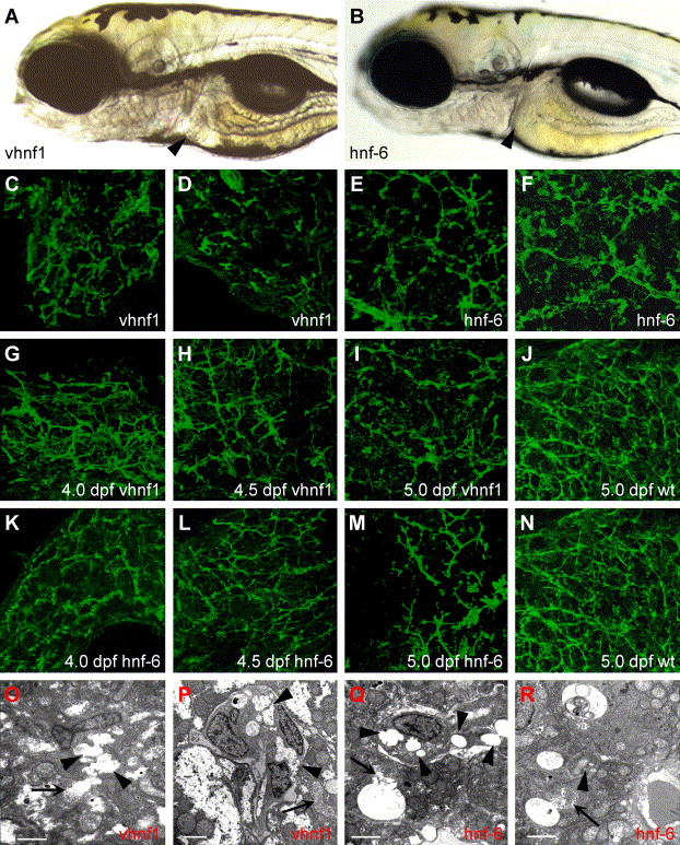
Fig. 10 Forced expression of hnf-6 or vhnf1 perturbs biliary development. (A?B) Left lateral views of 5 dpf larva injected with vhnf1 (A) or hnf-6 (B) mRNA. These larvae are virtually indistinguishable from WT larvae. Liver size is normal in both larvae (arrowheads). (C?N) Confocal projections through the liver of vhnf1 (C, D, G?I) and hnf-6 (E, F, K?M) RNA-injected larvae processed for cytokeratin IHC. Forced expression of either gene disrupts biliary development of 5 dpf larvae (C?F, I, and M). Few normal bile ducts are seen in most RNA-injected larvae compared with WT (J and N). (G?I, K?M) Biliary morphology of 4 dpf-5 dpf vhnf1 RNA (G?I) and hnf-6 RNA (K?M) larvae. Bile duct development appears abnormal at all developmental time points when compared with 5 dpf WT (J and N) and at earlier stages (Fig. 4G?I). (O?R) Liver ultrastructure in 5dpf vhnf1 (O and P) and hnf-6 (Q and R) RNA-injected larvae. There is evidence of cholestasis in these larvae. Thin arrows point to canaliculi; these are dilated in both sets of RNA-injected larvae. In hnf-6 RNA larvae (Q and R), bile has overflowed into the hepatocyte cytoplasm. Arrowheads in these figures point to dilated ducts. Prominent cholestatic debris is present within duct lumen in (P and R). Scale bars = 2 μm.
Reprinted from Developmental Biology, 274(2), Matthews, R.P., Lorent, K., Russo, P., and Pack, M., The zebrafish onecut gene hnf-6 functions in an evolutionarily conserved genetic pathway that regulates vertebrate biliary development, 245-259, Copyright (2004) with permission from Elsevier. Full text @ Dev. Biol.

