
|
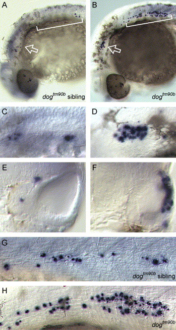
Fig. 6 Increased apoptotic cell death in dogtm90b. Whole mount visualization of TUNEL-positive cells (blue) in wild type (A, C, E, and G) and dogtm90b mutant (B, D, F, and H) embryos. (A and B) Lateral views of wild-type sibling (A) and dogtm90b (B) embryos at 28 hpf. In mutant embryos, increased numbers of TUNEL-positive cells are seen in the region of the otocyst (arrow) and migrating primordium of the posterior lateral line (bracket). (C and D). Higher magnification view of otocysts in wild-type sibling (C) and dogtm90b (D) embryos. (E and F) Transverse sections through the otic vesicles of embryos shown in panels A and B. Note that TUNEL-positive cells are located within the lateral otocyst wall of dogtm90b embryo. (G and H) Higher magnification view of TUNEL-positive cells in the migrating primordium of the posterior lateral line in wild-type sibling (G) and dogtm90b (H) embryos shown in A and B, respectively. Panels E and F: dorsal to top and lateral to right. All other panels: lateral views with dorsal to top and anterior to left.
Reprinted from Developmental Biology, 277(1), Kozlowski, D.J., Whitfield, T.T., Hukriede, N.A., Lam, W.K., and Weinberg, E.S., The zebrafish dog-eared mutation disrupts eya1, a gene required for cell survival and differentiation in the inner ear and lateral line, 27-41, Copyright (2005) with permission from Elsevier. Full text @ Dev. Biol.