
|
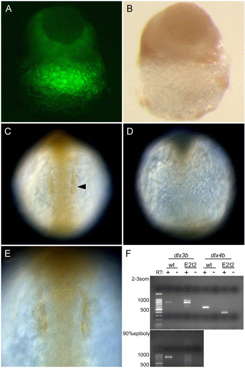
Fig. 1 Knockdown of dlx proteins in embryos injected with dlx3b/dlx4b-ATG-MOs and dlx3b/dlx4b-E2I2-MOs. The knockdown by the ATG-MOs was revealed by immunostaining with anti-Dll antibody (A?E). (A and B) Late blastula embryos. (C?E) 6-somite-stage embryos. (C and E) Wild-type embryos. (A, B, D) ATG-MOs-injected embryos. (A and B) Same images of a blastula. (A) Fluorescence image revealing MOs + LFD, and (B) bright field. The dispersion of MOs and anti-Dll immunoreactivity are complementary in the epiblast. Lateral views. (C and D) Anti-Dll antibody immunoreactivity in the otic placodal primordium (arrowhead) is absent in MOs-injected embryos. Dorsal views, anterior is to the top. (E) The magnification view of the otic placodal primordium in (C). The knockdown by the E2I2-MOs was revealed by RT-PCR followed by gel electrophoresis (F). (F) Wild type (wt) as compared to E2I2 MO (E2I2) dlx3b and dlx4b RT-PCR products. In dlx3b RT-PCR, at 2?3-somite stage, the amplification of cDNA from wild-type embryos produced an 810-bp band, and cDNA from E2I2-MOs-injected embryos produced three bands of 630, 810, and 900-bp in size. At 90% epiboly, in E2I2-MOs-injected embryos, the 810-bp band was faint. In dlx4b RT-PCR, at 2- to 3-somite stage, the amplification of cDNA from wild-type embryos produced a 580-bp band, and cDNA from E2I2-MO-injected embryos produced a 390-bp band. Negative-control reactions run in parallel, where reverse transcriptase was omitted in the cDNA synthesis reactions (RT-).
Reprinted from Developmental Biology, 276(2), Kaji, T., and Artinger, K.B., dlx3b and dlx4b function in the development of Rohon-Beard sensory neurons and trigeminal placode in the zebrafish neurula, 523-540, Copyright (2004) with permission from Elsevier. Full text @ Dev. Biol.