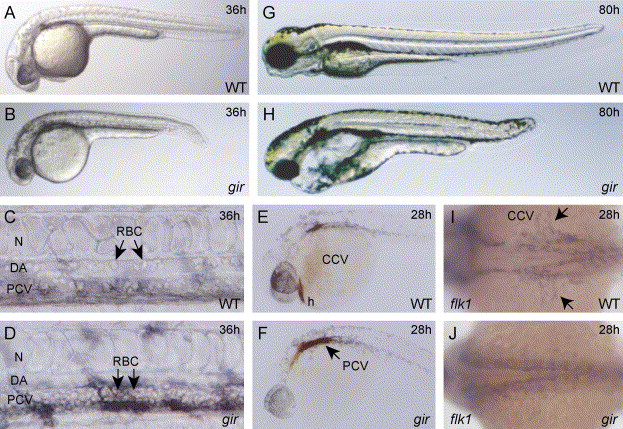
Fig. 1 Phenotypes of the gir mutant. (A and B) Morphology of the wild type (A) and gir mutant (B) embryos at 36 hpf. The gir mutant embryo exhibits a shortened tail. (C and D) Blood cells in dorsal aorta and posterior cardinal vein at 36 hpf. Circulating blood cells are evident in both dorsal aorta and posterior cardinal vein in wild-type embryos (C), whereas they are absent in the dorsal aorta and have accumulated in the posterior cardinal vein in gir mutant embryos (D). Photographs were taken after the embryos had been cooled on ice to make the circulation slower. (E and F) o-dianisidine staining of blood cells in wild type (E) and gir mutant (F) embryos at 28 hpf. Blood cells are seen in the common cardinal vein and heart in the wild-type embryo (E), whereas these cells have accumulated in the posterior cardinal vein and have failed to circulate in the gir mutant embryo (F). (G and H) Morphology of the wild-type (G) and gir mutant (H) embryos at 80 hpf. The gir mutant shows severe edema. (I and J) Expression of flk1 at 28 hpf. flk1 is expressed in the blood vessels including the common cardinal vein in wild-type embryo (I), whereas its expression is absent in the common cardinal vein of the gir mutant embryo (J). The arrows indicate the common cardinal vein. CCV, common cardinal vein; DA, dorsal aorta; h, heart; N, notochord; PCV, posterior cardinal vein; RBC, red blood cells.
Reprinted from Developmental Biology, 278(2), Emoto, Y., Wada, H., Okamoto, H., Kudo, A., and Imai, Y., Retinoic acid-metabolizing enzyme Cyp26a1 is essential for determining territories of hindbrain and spinal cord in zebrafish, 415-427, Copyright (2005) with permission from Elsevier. Full text @ Dev. Biol.

