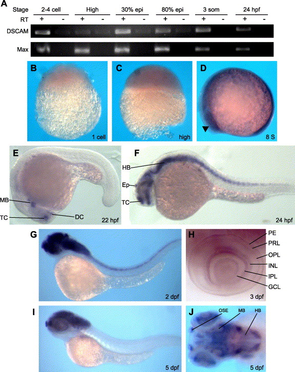
Fig. 2 Expression of zebrafish dscam during early embryogenesis. (A) RT-PCR of dscam at various developmental stages within the first 24 h. Max is used as loading control. Whole mount in situ hybridization of wild-type embryos at (B) 1 cell, (C) high, (D) 8 somite, (E) 22 hpf, (F) 24 hpf, (G) 2 dpf, (H) high magnification of 3 dpf eye, (I) 5 dpf, and (J) anterior?dorsal view at 5 dpf. B?D show expression throughout the early embryo. Arrowhead in D indicates the presumptive eye field. (E) 22 hpf embryo shows initial dscam discrete expression within midbrain, diencephalon, and telencephalon. (F) 24 hpf embryo shows expanded dscam expression and new expression in hindbrain and spinal cord. (G) 2 dpf embryo shows expanding brain and spinal cord expression. (H) Multiple layers of the laminated eye show strong dscam expression, particularly IPL and INL. (I) 5 dpf embryo shows expression only in the brain. (J) High magnification dorsal view of the brain shows dscam expression is expressed discretely throughout. PE, pigmented epithelium; PRL, photoreceptor layer; OPL, outer plexiform layer; INL, inner nerve layer; IPL, inner plexiform layer; GCL, ganglion cell layer; Ep, epiphysis; DC, diencephalon; HB, hindbrain; MB, midbrain; TC, telencephalon; OSE, olfactory sensory epithelium.
Reprinted from Developmental Biology, 279(1), Yimlamai, D., Konnikova, L., Moss, L.G., and Jay, D.G., The zebrafish down syndrome cell adhesion molecule is involved in cell movement during embryogenesis, 44-57, Copyright (2005) with permission from Elsevier. Full text @ Dev. Biol.

